J Korean Soc Menopause.
2011 Dec;17(3):174-177. 10.6118/jksm.2011.17.3.174.
Cervical Endometriosis in a Post-menopausal Woman: A Case Report
- Affiliations
-
- 1Department of Obstetrics and Gynecology, College of Medicine, Soonchunhyang University Bucheon Hospital, Bucheon, Korea. heeobgy@schmc.ac.kr
- 2Department of Pathology, College of Medicine, Soonchunhyang University Bucheon Hospital, Bucheon, Korea.
- KMID: 1444807
- DOI: http://doi.org/10.6118/jksm.2011.17.3.174
Abstract
- Cervical endometriosis is defined as the presence of endometrial glands and stroma at the cervix. This is rare and sometimes asymptomatic. Most of these are diagnosed by incidental findings within histopathology. As the presence of cytological features do not guarantee the presence of cervical endometriosis, it is difficult to diagnose this disorder prior to surgery. We recently encountered a case of cervical endometriosis in a post-menopausal woman who was not receiving hormone therapy. As a reminder to clinicians about this neglected issue, we report a case of cervical endometriosis with a literature review.
Keyword
Figure
-
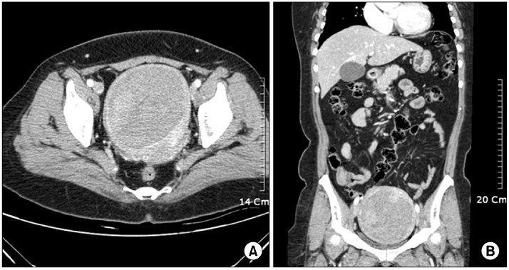
Fig. 1 (A) Axial view at arterial phase. (B) Coronal view at arterial phase. 11.0 × 9.0 × 10.2 cm sized heterogeneous enhancing mass was located at fundus and body portion of uterus.

Fig. 2 (A~C) × 10, × 40, × 200 H&E. Low magnification show several small and large endometrial glands with surrounding stroma admixed with endocercvical glands at the cervix. (D) Endometrial stromal cells exhibits CD10 expression.
Reference
-
1. Kim TH, Lee HH. Hemoperitoneum during pregnancy with endometriosis; report of four cases. Iran J Reprod Med. 2010. 8:90–93.2. Iwase A, Goto M, Kurotsuchi S, Harata T, Kaseki S, Kikkawa F. Successful management of a massive hemorrhage due to rupture of cystic cervical endometriosis by a loop electrosurgical excision procedure. Fertil Steril. 2008. 89:991e13–e15.3. Phadnis SV, Doshi JS, Ogunnaike O, Coady A, Padwick M, Sanusi FA. Cervical endometriosis: a diagnostic and management dilemma. Arch Gynecol Obstet. 2005. 272:289–293.4. Wong FW, Lim CE, Karia S, Santos L. Cervical endometriosis: case series and review of literature. J Obstet Gynaecol Res. 2010. 36:916–919.5. Yokota N, Yoshida H, Sakakibara H, Inayama Y, Hirahara F. A severe vaginal hemorrhage caused by cervical endometriosis. Am J Obstet Gynecol. 2008. 199:e12–e13.6. Chang SH, Maddox WA. Adenocarcinoma arising within cervical endometriosis and invading the adjacent vagina. Am J Obstet Gynecol. 1971. 110:1015–1017.7. Noda K, Kimura K, Ikeda M, Teshima K. Studies on the histogenesis of cervical adenocarcinoma. Int J Gynecol Pathol. 1983. 1:336–346.8. Veiga-Ferreira MM, Leiman G, Dunbar F, Margolius KA. Cervical endometriosis: facilitated diagnosis by fine needle aspiration cytologic testing. Am J Obstet Gynecol. 1987. 157:849–856.9. Kim TH, Lee HH, Chung SH, Kwak JJ, Park HS. Endometriosis detected in postmenopausal women not receiving menopausal hormone therapy: two case reports. J Korean Soc Menopause. 2010. 16:176–180.10. Kim TH, Lee HH, Chung SH, Kwak JJ, Lee BI, Hong YP. Serous adenocarcinoma arising from ovarian endometriosis after menopause. Korean J Obstet Gynecol. 2010. 53:365–370.11. Baker PM, Clement PB, Bell DA, Young RH. Superficial endometriosis of the uterine cervix: a report of 20 cases of a process that may be confused with endocervical glandular dysplasia or adenocarcinoma in situ. Int J Gynecol Pathol. 1999. 18:198–205.12. Szyfelbein WM, Baker PM, Bell DA. Superficial endometriosis of the cervix: A source of abnormal glandular cells on cervicovaginal smears. Diagn Cytopathol. 2004. 30:88–91.13. Hoang NM, Smadja A, Orcel L. Endometriosis of the uterine cervix. A hypothesis on its histogenesis. J Gynecol Obstet Biol Reprod (Paris). 1987. 16:587–593.
- Full Text Links
-
- Actions
-
Cited
- CITED
-
- Close
- Share
-
- Similar articles
-
- Endometriosis in a Postmenopausal Woman on Hormonal Replacement Therapy
- Postmenopausal Spontaneous Umbilical Endometriosis: A Case Report
- Extrauterine Undifferentiated Uterine Sarcoma Arising from Bladder Endometriosis in a Postmenopausal Woman: A Case Report
- A Case of Cervical Endometriosis with Cystic Change
- Ureteral Endometriosis
