J Korean Soc Radiol.
2011 Jun;64(6):599-602. 10.3348/jksr.2011.64.6.599.
Radiologic Findings of an Angioleiomyoma of the Finger: A Case Report
- Affiliations
-
- 1Department of Diagnostic Radiology, Seoul Paik Hospital, InJe University College of Medicine, Korea. jcshim@unitel.co.kr
- 2Department of Orthopedic Surgery, Seoul Paik Hospital, InJe University College of Medicine, Korea.
- 3Department of Pathology, Seoul Paik Hospital, InJe University College of Medicine, Korea.
- KMID: 1443511
- DOI: http://doi.org/10.3348/jksr.2011.64.6.599
Abstract
- Angioleiomyomas are a rare benign smooth muscle tumor arising from vessel walls. Although angioleiomyomas are most frequently reported in the lower extremities and in middle-aged female patients, they can be found throughout the body in male and female adults of all ages. We report a rare case of an angioleiomyoma of the left 3rd digit in a 31-year-old man, which appeared as a small, well defined mass with multiple vascular structures on Doppler sonogram and MRI. The tumor was diagnosed by pathology as an angioleiomyoma. Although angioleiomyomas are relatively infrequent, they should be considered in the differential diagnosis when multiple tortuous vascular structures are seen within a well demarcated mass in extremities on Doppler sonogram and MRI.
MeSH Terms
Figure
-
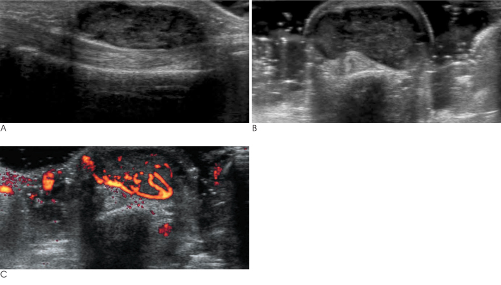
Fig. 1 Longitudinal (A) and transverse (B) gray scale sonograms show a well defined heterogeneous low echoic mass just above the flexor tendon of the 3rd finger. Power Doppler sonogram (C) demonstrates multiple vascular channels within the tumor.
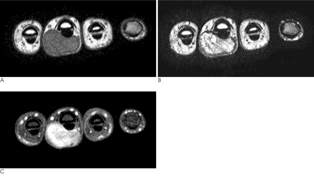
Fig. 2 The axial T1 weighted (A) and T2 weighted (B) images demonstrate well defined a mass with a thin capsule; curvilinear low signal structures are also noted within the mass (arrows in A and B). Gadolinium-enhanced T1 weighted image (C) demonstrate strong enhancement, especially peripherally.
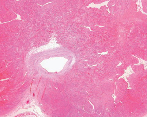
Fig. 3 Photomicroscopic image (H & E ×10) demonstrates compact smooth muscle cells surrounding the vessel wall. Multiple small sized split-like vascular channels are also noted.
Reference
-
1. Hachisuga T, Hashimoto H, Enjoji M. Angioleiomyoma: a clinicopathologic reappraisal of 562 cases. Cancer. 1984; 54:126–130.2. Freedman AM, Meland NB. Angioleiomyomas of the extremities: report of a case and review of the Mayo Clinic experience. Plast Reconstr Surg. 1989; 83:328–331.3. Morimoto N. Angioleiomyoma: a clinicopathologic study. Med J Kagoshima Univ. 1973; 24:663–683.4. Hwang JW, Ahn JM, Kang HS, Suh JS, Kim SM, Seo JW. Vascular leiomyoma of an extremity: MR imaging-pathology correlation. AJR Am J Roentgenol. 1998; 171:981–985.5. Yoo HJ, Choi JA, Chung JH, Oh JH, Lee GK, Choi JY, et al. Angioleiomyoma in soft tissue of extremities: MRI findings. AJR Am J Roentgenol. 2009; 192:291–294.6. Kinoshita T, Ishii K, Abe Y, NAganuma H. Angiomyoma of the lower extremity: MR findings. Skeletal Radiol. 1997; 26:443–445.7. Theumann NH, Bittoun J, Goettmann S, Le Viet D, Chevrot A, Drapé JL. Hemangiomas of the fingers: MR imaging evaluation. Radiology. 2001; 218:841–847.
