J Korean Soc Radiol.
2011 Aug;65(2):167-170. 10.3348/jksr.2011.65.2.167.
Synovial Sarcoma in the Rectovesical Space: A Case Report
- Affiliations
-
- 1Department of Radiology, Chungbuk National University Hospital, Cheongju, Korea. sircircle@chungbuk.ac.kr
- 2Department of Pathology, Chungbuk National University Hospital, Cheongju, Korea.
- KMID: 1443481
- DOI: http://doi.org/10.3348/jksr.2011.65.2.167
Abstract
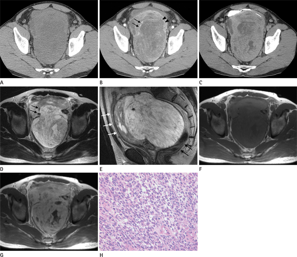
- Synovial sarcoma is an uncommon soft tissue malignancy usually arising in the extremities of young adults. Synovial sarcomas at unusual anatomic locations have been reported; however, to the best of our knowledge, there are no reports on primary synovial sarcoma in the rectovesical space. Here, we describe the radiologic findings of primary synovial sarcoma in the rectovesical space and review relevant literature.
Figure
Reference
-
1. Murphey MD, Gibson MS, Jennings BT, Crespo-Rodríguez AM, Fanburg-Smith J, Gajewski DA. From the archives of the AFIP: Imaging of synovial sarcoma with radiologicpathologic correlation. Radiographics. 2006; 26:1543–1565.2. Ulusan S, Kizilkilic O, Yildirim T, Hurcan C, Bal N, Nursal TZ. Radiological findings of primary retroperitoneal synovial sarcoma. Br J Radiol. 2005; 78:166–169.3. Chatzipantelis P, Kafiri G. Retroperitoneal synovial sarcoma: a clinicopathological study of 6 cases. J BUON. 2008; 13:211–216.4. Janke WH, Block MA. Chronic retroperitoneal pelvic abscesses. Arch Surg. 1965; 90:380–384.5. Tosaka A, Yamazaki A, Hirokawa M, Matsushita K, Asakura S. [A bulky mass non-Hodgkin's lymphoma with dysuria in the rectovesical space]. Hinyokika Kiyo. 1990; 36:701–705.6. Wilson RG. Dermoid cyst of the rectovesical space: report of a case. Dis Colon Rectum. 1973; 16:530–531.7. Morton MJ, Berquist TH, McLeod RA, Unni KK, Sim FH. MR imaging of synovial sarcoma. AJR Am J Roentgenol. 1991; 156:337–340.8. Tateishi U, Hasegawa T, Beppu Y, Satake M, Moriyama N. Synovial sarcoma of the soft tissues: prognostic significance of imaging features. J Comput Assist Tomogr. 2004; 28:140–148.9. Jones BC, Sundaram M, Kransdorf MJ. Synovial sarcoma: MR imaging findings in 34 patients. AJR Am J Roentgenol. 1993; 161:827–830.10. Nishimura H, Zhang Y, Ohkuma K, Uchida M, Hayabuchi N, Sun S. MR imaging of soft-tissue masses of the extraperitoneal spaces. Radiographics. 2001; 21:1141–1154.


