A Case of Novel Influenza A (H1N1) Virus Pneumonia Complicated Pnemomediastinum and Subcutenous Emphysema
- Affiliations
-
- 1Division of Pulmonology, Department of Internal Medicine, Gachon University Gil Hospital, Incheon, Korea. allergy21@hotmail.com
- 2Department of Laboratory Medicine, Gachon University Gil Hospital, Incheon, Korea.
- KMID: 1442866
- DOI: http://doi.org/10.4046/trd.2011.70.2.155
Abstract
- Recently, a novel influenza A (H1N1) has been recognized as the cause of a worldwide respiratory infection outbreak. Although the symptoms of a novel influenza A (H1N1) are usually mild, the disease can cause severe illness and death. A complication of novel influenza A (H1N1) is pneumomediastinum, a rarely reported condition. We report a case of influenza A (H1N1) complicating pneumomediastinum with subcutaneous emphysema, which had initially presented with blood tinged sputum and chest pain. In addition, we demonstrate bronchoalveolar lavage in influenza A (H1N1).
MeSH Terms
Figure
-

Figure 1 Chest radiograph on admission showed pneumomediastinum and peribronchial haziness in both lungs (A~C).

Figure 2 Neck radiograph on admission showed subcutaneous emphysema, neck and upper thorax (A, B).

Figure 3 High resolution computed tomography of chest on admission. (A) Showed subcutaneous inter-, intra-muscular emphysema in scan covered lower neck. (B~D) Peribronchial ground grass opacity in both lower lobe, right middle lobe, superior and inferior lingular segment of left upper lobe, posterior segment of right upper lobe.
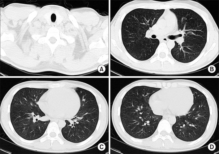
Figure 4 High resolution computed tomography of chest after 2 weeks. (A) Disappeared subcutaneous inter-, intra-muscular emphysema in scan covered lower neck. (B~D) Disappeared pneumomediastinum and improved peribronchial ground grass opacity in both lower lobe, right middle lobe, superior and inferior lingular segment of left upper lobe, posterior segment of right upper lobe.
Reference
-
1. Schnitzler SU, Schnitzler P. An update on swine-origin influenza virus A/H1N1: a review. Virus Genes. 2009. 39:279–292.2. Centers for Disease Control and Prevention (CDC). Hospitalized patients with novel influenza A (H1N1) virus infection - California, April-May, 2009. MMWR Morb Mortal Wkly Rep. 2009. 58:536–541.3. ANZIC Influenza Investigators. Webb SA, Pettilä V, Seppelt I, Bellomo R, Bailey M, et al. Critical care services and 2009 H1N1 influenza in Australia and New Zealand. N Engl J Med. 2009. 361:1925–1934.4. Bullaro FM, Bartoletti SC. Spontaneous pneumomediastinum in children: a literature review. Pediatr Emerg Care. 2007. 23:28–30.5. Newcomb AE, Clarke CP. Spontaneous pneumomediastinum: a benign curiosity or a significant problem? Chest. 2005. 128:3298–3302.6. Hasegawa M, Hashimoto K, Morozumi M, Ubukata K, Takahashi T, Inamo Y. Spontaneous pneumomediastinum complicating pneumonia in children infected with the 2009 pandemic influenza A (H1N1) virus. Clin Microbiol Infect. 2010. 16:195–199.7. Yun TJ, Kwon GJ, Oh MK, Woo SK, Park SH, Choi SH, et al. Radiological and Clinical Characteristics of a Military Outbreak of Pandemic H1N1 2009 Influenza Virus Infection. Korean J Radiol. 2010. 11:417–424.8. Choi WJ, Kim WY, Kim SH, Oh BJ, Kim W, Lim KS, et al. Clinical characteristics of pneumonia in hospitalized patients with novel influenza A (H1N1) in Korea. Scand J Infect Dis. 2010. 42:311–314.9. Reynolds HY. Bronchoalveolar lavage. Am Rev Respir Dis. 1987. 135:250–263.10. Magnan A, Mege JL, Reynaud M, Thomas P, Capo C, Garbe L, et al. Monitoring of alveolar macrophage production of tumor necrosis factor-alpha and interleukin-6 in lung transplant recipients. Marseille and Montreal Lung Transplantation Group. Am J Respir Crit Care Med. 1994. 150:684–689.
- Full Text Links
-
- Actions
-
Cited
- CITED
-
- Close
- Share
-
- Similar articles
-
- Spontaneous Pneumomediastinum, Pneumothorax, and Subcutaneous Emphysema Complicating H1N1 Virus Infection
- Two Cases of Spontaneous Pneumomediastinum Complicating Viral Pneumonia Caused by Influenza A Virus, (H1N1 Subtype): A Case Report
- Influenza Associated Pneumonia
- A Case of Pseudomembranous Tracheobronchitis Complicated by Coinfection of 2009 Pandemic Influenza A/H1N1 and Staphylococcus aureus
- 2009 H1N1 influenza virus infection and necrotizing pneumonia treated with extracorporeal membrane oxygenation
