Abnormal Positioning of Multiple Abdominal Organs with Anomalous Direct Drainage of Hepatic Vein into the Right Atrium in a Post-Operative Omphalocele Patient: A Case Report
- Affiliations
-
- 1Department of Radiology, Chungbuk National University Hospital, Cheongju, Korea. sircircle@chungbuk.ac.kr
- KMID: 1439544
- DOI: http://doi.org/10.3348/jksr.2012.67.4.293
Abstract
- An omphalocele is a rare congenital anomaly in which the infant's intestines protrude through the navel. Additional anomalies that are associated with omphalocele remain present in as many as 50% of cases, and these anomalies vary greatly from patient to patient. However, the persistent anomalies or abnormal position of the abdominal organs in post-operative omphalocele patients have not reported previously. Herein, we report the case of an omphalocele patient with abnormal positioning of the liver, spleen and both kidneys, as well as abnormal drainage of the hepatic vein into the right atrium, which was found during a routine, postoperative follow-up computed tomography scan.
MeSH Terms
Figure
-
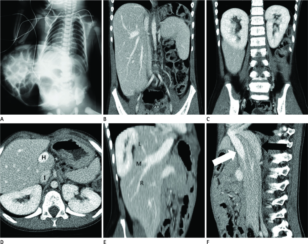
Fig. 1 A 6-year-old girl with a temporary silo placed because of a large omphalocele. A. Initial infantogram on the first day of life. It shows multiple bowel loop herniations into a membrane-covered sac. Radiopaque portion within the sac is thought to represent portions of herniated liver and spleen. B. Follow-up abdomen and pelvis computed tomography 6 years later reveals a craniocaudal liver position. C. Both kidneys are located in just beneath the diaphragm. D. Liver, spleen and both kidneys are shown in the same level, just beneath the diaphragm. The hepatic vein (H) and IVC (I) run to the right atrium, independently. E. Three major hepatic vein show within the liver to form a common trunk. F. Common hepatic vein (white arrow) directly drains into the right atrium, instead of conjoining with the IVC (black arrow). The IVC also drains into the right atrium, independently. Note.-IVC = inferior vena cava, L = left, M = middle, R = right hepatic vein
Reference
-
1. Antoniou EE, Matsuoka S, Mori K, Hayabuchi Y, Kuroda Y. Anomalous inferior vena cava in association with omphalocele: a case report. Pediatr Radiol. 1995; 25:265–266.2. Christison-Lagay ER, Kelleher CM, Langer JC. Neonatal abdominal wall defects. Semin Fetal Neonatal Med. 2011; 16:164–172.3. Onouchi Z, Ootsuka T, Otabe E, Sasagawa H, Takada Y. A case of the omphalocele. The discussion about the frequent combination of the cardiac malformation, and the observation at the early non-cyanotic period of Fallot's tetralogy. Jpn Heart J. 1975; 16:211–220.4. Zaccara A, Iacobelli BD, La Sala E, Calzolari A, Turchetta A, Orazi C, et al. Sonographic biometry of liver and spleen size long after closure of abdominal wall defects. Eur J Pediatr. 2003; 162:490–492.5. van Eijck FC, Klein WM, Boetes C, Aronson DC, Wijnen RM. Has the liver and other visceral organs migrated to its normal position in children with giant omphalocele? A follow-up study with ultrasonography. Eur J Pediatr. 2010; 169:563–567.6. D'Cruz IA, Smith KA. Echocardiographic appearance of direct drainage of hepatic vein into right atrium in adults. Echocardiography. 1995; 12:465–468.7. Waldman JD, Fellows KE, Paul MH, Muster AJ. Angulation of the inferior vena cava-right atrial junction in children with repaired omphalocele. Pediatr Radiol. 1977; 5:142–144.
- Full Text Links
-
- Actions
-
Cited
- CITED
-
- Close
- Share
-
- Similar articles
-
- Hepatic Venous Return in Atrial Isomerism Evaluated by MR
- Levoatriocardinal Vein Combined with Pulmonary Venous Varix Mimicking Arteriovenous Malformations: A Case Report
- Anomalous Coronary Sinus Drainage Into the Left Atrium: A Case Report and Brief Review
- Modified Extracardiac Fontan Procedure in a Univentricular Heart with Separate Hepatic and Inferior Vena Caval Drainage
- A Case of Surgically Corrected-Combined form of Total Anomalous Pulmonary Venous Return
