J Korean Bone Joint Tumor Soc.
2012 Jun;18(1):41-44. 10.5292/jkbjts.2012.18.1.41.
Multiple Glomus Tumor in Brachial Plexus: A Case Report
- Affiliations
-
- 1Department of Orthopedic Surgery, School of Medicine, Kyung Hee University, Seoul, Korea. dukech@khmc.or.kr
- KMID: 1438411
- DOI: http://doi.org/10.5292/jkbjts.2012.18.1.41
Abstract
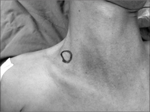
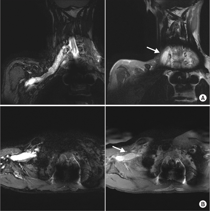
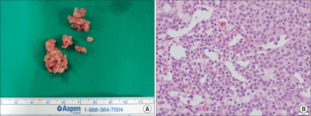
- Glomus tumor is a kind of vascular tumor that arises from the glomus body, which regulates skin temperature and is placed in the skin and the subcutaneous area. It is a benign tumor that usually presents in the subungal area. It is relatively common in areas other than the fingers, but its occurrence in peripheral nerves is known to be comparatively rare. We report our experience with a case of glomus tumor arising from the brachial plexus, a rare site of occurrence for glomus tumors.
Keyword
Figure
Reference
-
1. Carroll RE, Berman AT. Glomus tumors of the hand: review of the literature and report on twenty-eight cases. J Bone Joint Surg Am. 1972. 54:691–703.2. Schiefer TK, Parker WL, Anakwenze OA, Amadio PC, Inwards CY, Spinner RJ. Extradigital glomus tumors: a 20-year experience. Mayo Clin Proc. 2006. 81:1337–1344.3. Takei TR, Nalebuff EA. Extradigital glomus tumour. J Hand Surg Br. 1995. 20:409–412.4. Kim SH, Suh HS, Choi JH, Sung KJ, Moon KC, Koh JK. Glomus tumor: a clinical and histopathologic analysis of 17 cases. Ann Dermatol. 2000. 12:95–101.5. Kim PT, Park IH, Oh SH. Ultrasonography of a subungual glomus tumor -cases report-. J Korean Soc Surg Hand. 1999. 4:117–120.6. Maxwell GP, Curtis RM, Wilgis EF. Multiple digital glomus tumors. J Hand Surg Am. 1979. 4:363–367.7. Scheithauer BW, Rodriguez FJ, Spinner RJ, et al. Glomus tumor and glomangioma of the nerve. Report of two cases. J Neurosurg. 2008. 108:348–356.


