J Korean Assoc Oral Maxillofac Surg.
2012 Oct;38(5):271-275. 10.5125/jkaoms.2012.38.5.271.
Three-dimensional finite element analysis of the stress distribution and displacement in different fixation methods of bilateral sagittal split ramus osteotomy
- Affiliations
-
- 1Department of Oral and Maxillofacial Surgery, St. Paul's Hospital, The Catholic University of Korea, College of Medicine, Seoul, Korea.
- 2Department of Oral and Maxillofacial Surgery, The Catholic University of Korea, College of Medicine, Seoul, Korea.
- 3Department of Oral and Maxillofacial Surgery, Seoul St. Mary's Hospital, The Catholic University of Korea, College of Medicine, Seoul, Korea. jupark@catholic.ac.kr
- 4Department of Oral and Maxillofacial Surgery, Uijeongbu St. Mary's Hospital, The Catholic University of Korea, College of Medicine, Uijeongbu, Korea.
- 5Department of Mechanical Engineering, Myongji University, Seoul, Korea.
- KMID: 1434327
- DOI: http://doi.org/10.5125/jkaoms.2012.38.5.271
Abstract
OBJECTIVES
This study evaluated a range of fixation methods to determine which is best for the postoperative stabilization of a mandibular osteotomy using three-dimensional finite element analysis of the stress distribution on the plate, screw and surrounding bone and displacement of the lower incisors.
MATERIALS AND METHODS
The model was generated using the synthetic skull scan data, and the surface model was changed to a solid model using software. Bilateral sagittal split ramus osteotomy was performed using the program, and 8 different types of fixation methods were evaluated. A vertical load of 10 N was applied to the occlusal surface of the first molar.
RESULTS
In the case of bicortical screws, von-Mises stress on the screws and screw hole and deflection of the lower central incisor were minimal in type 2 (inverted L pattern with 3 bicortical repositioning screws). In the case of plates, von-Mises stress was minimal in type 8 (fixation 5 mm above the inferior border of the mandible with 1 metal plate and 4 monocortical screws), and deflection of the lower central incisor was minimal in types 6 (fixation 5 mm below the superior border of the mandible with 1 metal plate and 4 monocortical screws) and 7 (fixation 12 mm below the superior border of the mandible with 1 metal plate and 4 monocortical screws).
CONCLUSION
Types 2 and 6 fixation methods provide better stability than the others.
MeSH Terms
Figure
-
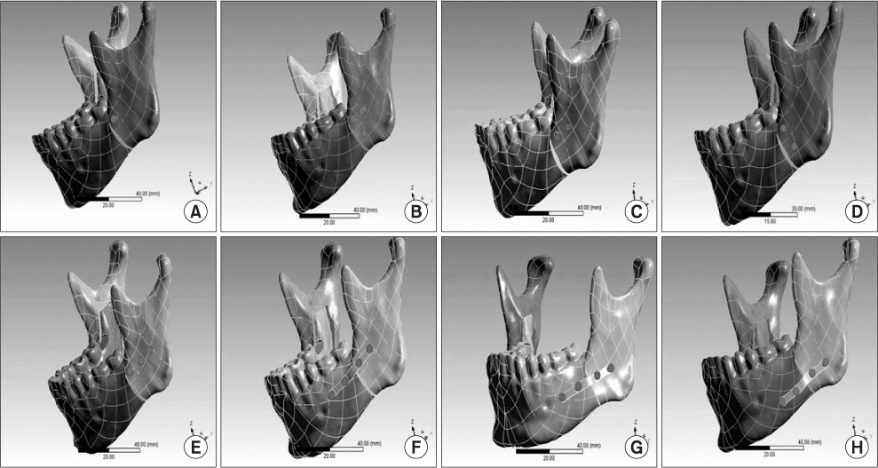
Fig. 1 Finite element model. A. Type 1. B. Type 2. C. Type 3. D. Type 4. E. Type 5. F. Type 6. G. Type 7. H. Type 8.
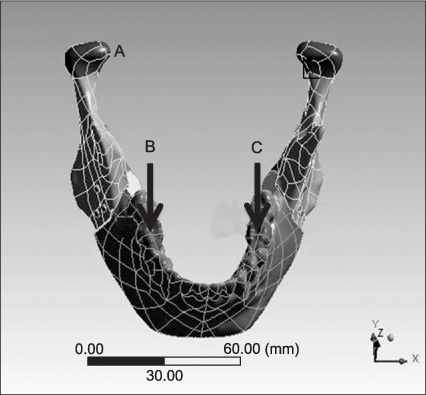
Fig. 2 Boundary conditions of the finite element model of the mandible. (A: fixed, B: force, C: force)
Reference
-
1. Park JU. Orthognathic surgery. 2003. Seoul: Koonja Publishing;267–316.2. Wolford LM. The sagittal split ramus osteotomy as the preferred treatment for mandibular prognathism. J Oral Maxillofac Surg. 2000. 58:310–312.
Article3. Oguz Y, Uckan S, Ozden AU, Uckan E, Eser A. Stability of locking and conventional 2.0-mm miniplate/screw systems after sagittal split ramus osteotomy: finite element analysis. Oral Surg Oral Med Oral Pathol Oral Radiol Endod. 2009. 108:174–177.
Article4. Ueki K, Hashiba Y, Marukawa K, Alam S, Nakagawa K, Yamamoto E. Skeletal stability after mandibular setback surgery: bicortical fixation using a 2.0-mm locking plate system versus monocortical fixation using a nonlocking plate system. J Oral Maxillofac Surg. 2008. 66:900–904.
Article5. Brasileiro BF, Grempel RG, Ambrosano GM, Passeri LA. An in vitro evaluation of rigid internal fixation techniques for sagittal split ramus osteotomies: advancement surgery. J Oral Maxillofac Surg. 2009. 67:809–817.
Article6. Jafari A, Shetty KS, Kumar M. Study of stress distribution and displacement of various craniofacial structures following application of transverse orthopedic forces--a three-dimensional FEM study. Angle Orthod. 2003. 73:12–20.7. Holberg C. Effects of rapid maxillary expansion on the cranial base--an FEM-analysis. J Orofac Orthop. 2005. 66:54–66.
Article8. Proffit WR, Phillips C. Adaptations in lip posture and pressure following orthognathic surgery. Am J Orthod Dentofacial Orthop. 1988. 93:294–302.9. Nagai I, Tanaka N, Noguchi M, Suda Y, Sonoda T, Kohama G. Changes in occlusal state of patients with mandibular prognathism after orthognathic surgery: a pilot study. Br J Oral Maxillofac Surg. 2001. 39:429–433.
Article10. Jang HS, Kim YK. A clinical study on the relapse after bssro for the correction of mandibular prognathism. J Korean Assoc Oral Maxillofac Surg. 1996. 22:4–9.11. Erkmen E, Simsek B, Yucel E, Kurt A. Comparison of different fixation methods following sagittal split ramus osteotomies using three-dimensional finite elements analysis. Part 1: advancement surgery-posterior loading. Int J Oral Maxillofac Surg. 2005. 34:551–558.
Article12. Yoon OB, Kim YG. A study of von-Mises yield strength after mandibular sagittal split ramus osteotomy. J Korean Assoc Oral Maxillofac Surg. 2002. 28:196–200.13. Lindorf HH. Sagittal ramus osteotomy with tandem screw fixation. Technique and results. J Maxillofac Surg. 1986. 14:311–316.14. Haug RH, Barber JE, Punjabi AP. An in vitro comparison of the effect of number and pattern of positional screws on load resistance. J Oral Maxillofac Surg. 1999. 57:300–308.
Article15. Swift JQ. Mandibular advancement. Atlas Oral Maxillofac Surg Clin North Am. 1993. 1:17–27.
Article16. Anucul B, Waite PD, Lemons JE. In vitro strength analysis of sagittal split osteotomy fixation: noncompression monocortical plates versus bicortical position screws. J Oral Maxillofac Surg. 1992. 50:1295–1299.
Article17. Chuong CJ, Borotikar B, Schwartz-Dabney C, Sinn DP. Mechanical characteristics of the mandible after bilateral sagittal split ramus osteotomy: comparing 2 different fixation techniques. J Oral Maxillofac Surg. 2005. 63:68–76.
Article
- Full Text Links
-
- Actions
-
Cited
- CITED
-
- Close
- Share
-
- Similar articles
-
- A Study Of Von-Mises Yield Strength After Mandibular Sagittal Split Ramus Osteotomy
- A case report of hemifacial microsomia
- Comparative study of stability and relapse according to fixation method after bilateral sagittal split ramus osteotomies in mandibular prognathic patients
- Stability after Surgical Correction of Mandibular Prognathism Using Bilateral Sagittal Split Ramus Osteotomy with Rigid Fixation
- A 3-D finite element analysis on the mandibular movement pattern and stress distribution during symphyseal widening
