Korean J Clin Microbiol.
2012 Dec;15(4):147-150. 10.5145/KJCM.2012.15.4.147.
Lung Abscess and Bacteremia Caused by Neisseria flavescens and Streptococcus sanguis in Patient with Idiopathic Hypereosinophilic Syndrome
- Affiliations
-
- 1Department of Pediatrics, Pusan National University School of Medicine, Yangsan, Korea. sonofs@naver.com
- 2Department of Laboratory Medicine, Pusan National University School of Medicine, Yangsan, Korea.
- 3Department of Laboratory Medicine, Pusan National University Hospital, Busan, Korea.
- KMID: 1431812
- DOI: http://doi.org/10.5145/KJCM.2012.15.4.147
Abstract
- Neisseria flavescens has been rarely reported as a pathogen in the literature. We experienced a case of N. flavescens bacteremia and lung abscess co-infected with Streptococcus sanguis in patient with idiopathic hypereosinophilic syndrome. A 15-year-old boy was diagnosed with idiopathic hypereosinophilic syndrome complicated with pulmonary thromboembolism. He was given systemic steroids and thrombolytics. After 8 weeks of therapy, a lung abscess appeared on the plain chest radiograph. We treated him with empirical antibiotics and carried out surgical drainage. Two types of microorganisms were cultured from both blood and pus samples, obtained in the first day of hospitalization. Pus was aspirated from the lung abscess with an aseptic technique. Neisseria species and S. sanguis were identified using traditional methods. To confirm the identity of the Neisseria species, we conducted further testing using 16S ribosomal ribonucleic acid sequencing whereupon N. flavescens was identified. This is the first case report of pulmonary infection caused by N. flavescens. We suggest that N. flavescens may act as a pathogen.
MeSH Terms
Figure
-
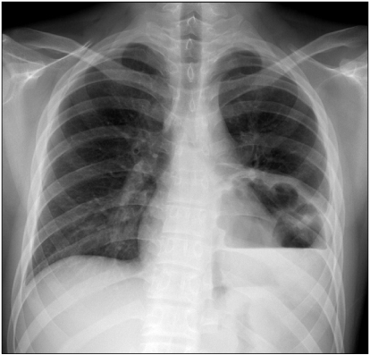
Fig. 1 Chest plain radiograph showing lung abscess.
Reference
-
1. Feder HM, Garibaldi RA. The significance of nongonococcal, nonmeningococcal Neisseria isolates from blood cultures. Rev Infect Dis. 1984. 6:181–188.2. Roufosse FE, Goldman M, Cogan E. Hypereosinophilic syndromes. Orphanet J Rare Dis. 2007. 2:37.3. Clinical and Laboratory Standards Institute. CLSI Document MM18-A. Interpretive criteria for identification of bacteria and fungi by DNA target sequencing: Approved guideline. 2008. Wayne, PA: Clinical and Laboratory Standards Institute.4. Wertlake PT, Williams TW Jr. Septicaemia caused by Neisseria flavescens. J Clin Pathol. 1968. 21:437–439.5. Coovadia YM. Neisseria flavescens septicaemia with meningitis. A case report. S Afr Med J. 1984. 66:308–309.6. Sinave CP, Ratzan KR. Infective endocarditis caused by Neisseria flavescens. Am J Med. 1987. 82:163–164.7. Szabo S, Lieberman JP, Lue YA. Unusual pathogens in narcoticssociated endocarditis. Rev Infect Dis. 1990. 12:412–415.8. Quintero Otero S, Rubio Quiñones F, Hernández Gonzalez A, Díaz Portillo J, García Martos P, Pantoja Rosso S. Septic shock caused by Neisseria flavescens. An Esp Pediatr. 1990. 33:64–65.9. Melican K, Dumenil G. Vascular colonization by Neisseria meningitidis. Curr Opin Microbiol. 2012. 15:50–56.10. Rosell A, Monsó E, Soler N, Torres F, Angrill J, Riise G, et al. Microbiologic determinants of exacerbation in chronic obstructive pulmonary disease. Arch Intern Med. 2005. 165:891–897.11. Cabrera-Rubio R, Garcia-Núñez M, Setó L, Antó JM, Moya A, Monsó E, et al. Microbiome diversity in the bronchial tracts of patients with chronic obstructive pulmonary disease. J Clin Microbiol. 2012. 50:3562–3568.12. Thorpe JE, Baughman RP, Frame PT, Wesseler TA, Staneck JL. Bronchoalveolar lavage for diagnosing acute bacterial pneumonia. J Infect Dis. 1987. 155:855–861.13. Kovalyk AP, Govda AV. Characteristics of microflora of laryngeal mucosa in healthy subjects and patients with cicatrical stenosis of the larynx. Vestn Otorinolaringol. 2010. 17–20.
- Full Text Links
-
- Actions
-
Cited
- CITED
-
- Close
- Share
-
- Similar articles
-
- Septic Knee Arthritis Caused by Streptococcus sanguis in a Patient with Osteoarthritis of the Knee
- Idiopathic Hypereosinophilic Syndrome Involving Thoracic Spine
- Anesthetic Management of a Patient with Idiopathic Hypereosinophilic Syndrome: A case report
- Pulmonary Involvement of Hypereosinophilic Syndrome: High-Resolution CT Findings in Three Patients
- A Case of Idiopathic Hypereosinophilic Syndrome Presenting Acute Pulmonary Edema
