Stress analysis of maxillary premolars with composite resin restoration of notch-shaped class V cavity and access cavity; Three-dimensional finite element study
- Affiliations
-
- 1Department of Conservative dentistry, School of Dentistry, Pusan National University, Korea. jeongkil@pusan.ac.kr
- 2Department of Mechanical design engineering, College of Engineering, Pusan National University, Korea.
- KMID: 1428468
- DOI: http://doi.org/10.5395/JKACD.2008.33.6.570
Abstract
- The purpose of this study was to investigate the distribution of tensile stress of canal obturated maxillary second premolar with access cavity and notch-shaped class V cavity restored with composite resin using a 3D finite element analysis. The tested groups were classified as 8 situations by only access cavity or access cavity with notch-shaped class VS cavity (S or N), loading condition (L1 or L2), and with or without glass ionomer cement base (R1 or R2). A static load of 500 N was applied at buccal and palatal cusps. Notch-shaped cavity and access cavity were filled microhybrid composite resin (Z100) with or without GIC base (Fuji II LC). The tensile stresses presented in the buccal cervical area, palatal cervical area and occlusal surface were analyzed using ANSYS. Tensile stress distributions were similar regardless of base. When the load was applied on the buccal cusp, excessive high tensile stress was concentrated around the loading point and along the central groove of occlusal surface. The tensile stress values of the tooth with class V cavity were slightly higher than that of the tooth without class V cavity. When the load was applied the palatal cusp, excessive high tensile stress was concentrated around the loading point and along the central groove of occlusal surface. The tensile stress values of the tooth without class V cavity were slightly higher than that of the tooth with class V cavity.
MeSH Terms
Figure
-

Figure 1 Schematic representation of restored endodontically treated maxillary premolars with access cavity only (SR1 and SR2) or access cavity and notch-shaped class V cavity (NR1 and NR2). Dark brown; Composite resin restoration. Light brown; Glass ionomer cement base.

Figure 2 Schematic diagram of loading points

Figure 3 The maximum principal stress distribution of buccal surface under Load 1.

Figure 4 The maximum principal stress distribution of buccal surface under Load 1

Figure 5 The maximum principal stress distribution of occlusal surface under Load 1.

Figure 6 The maximum principal stress distribution along the central groove of occlusal surface under Load 1.
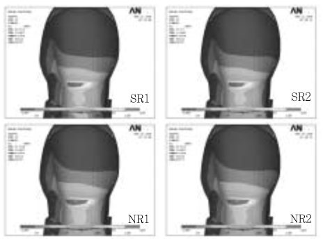
Figure 7 The maximum principal stress distribution of palatal surface under Load 1.
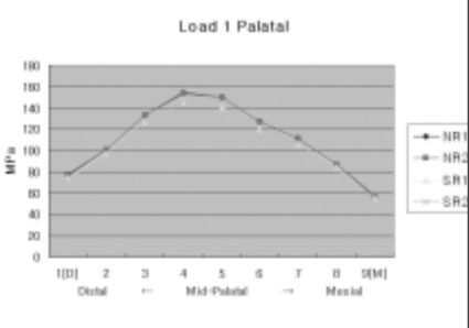
Figure 8 The maximum principal stress distribution of palatal surface under Load 1

Figure 9 The maximum principal stress distribution of buccal surface under Load 2.

Figure 10 The maximum principal stress distribution of buccal surface under Load 2.

Figure 11 The maximum principal stress distribution of occlusal surface under Load 2.

Figure 12 The maximum principal stress distribution along the central groove of occlusal surface under Load 2.

Figure 13 The maximum principal stress distribution of palatal surface under Load 2.

Figure 14 The maximum principal stress distribution of palatal surface under Load 2.
Reference
-
1. Reeh ES, Messer HH, Douglas WH. Reduction in tooth stiffness as a result of endodontic and restorative procedures. J Endod. 1989. 15:512–516.
Article2. Helfer AR, Melnick S, Schilder H. Determination of the moisture content of vital and pulpless teeth. Oral Surg Oral Med Oral Pathol. 1972. 34:661–670.
Article3. Panitvisai P, Messer HH. Cuspal deflection in molars in relation to endodontic and restorative procedures. J Endod. 1995. 21:57–61.
Article4. Lewinstein I, Grajower R. Root dentin hardness of endodontically treated teeth. J Endod. 1981. 7:421–422.
Article5. Fusayama T, Maeda T. Effect of pulpectomy on dentin hardness. J Dent Res. 1969. 48:452–460.
Article6. Pilo R, Cardash HS, Levin E, Assif D. Effect of core stiffness on the in vitro fracture of crowned, endodontically treated teeth. J Prosthet Dent. 2002. 88:302–306.
Article7. Gwinnett AJ. The morphologic relationship between dental resins and etched dentin. J Dent Res. 1977. 56:1155–1160.
Article8. Wolanek GA, Loushine RJ, Weller RN, Kimbrough WF, Volkmann KR. In vitro bacterial penetration of endodontically treated teeth coronally sealed with a dentin bonding agent. J Endod. 2001. 27:354–357.
Article9. Trope M, Langer I, Maltz D, Tronstad L. Resistance to fracture of restored endodontically treated premolars. Endod Dent Traumatol. 1986. 2:35–38.
Article10. Trope M, Tronstad L. Resistance to fracture of endodontically treated premolars restored with glass ionomer cement or acid etch composite resin. J Endod. 1991. 17:257–259.
Article11. Park JK, Hur B, Kim SK. Stress distribution of class V composite resin restorations: A three-dimensional finite element study. J Korean Acad Conserv Dent. 2008. 33:28–38.
Article12. Piotrowski BT, William B, Hancock . Examining the prevalence and characteristics of abfraction like cervical lesions in a population of U.S. veterans. J Am Dent Assoc. 2001. 132:1694–1701.
Article13. Park JK, Hur B, Kim SK. The influence of combining composite resins with different elastic modulus on the stress distribution of class V restoration : A three-dimensional finite element study. J Korean Acad Conserv Dent. 2008. 33:184–197.
Article14. Telles D, Pegoraro LF, Pereira JC. Prevalence of noncarions cervical lesions and their relation to occlusal aspects: a clinical study. J Esthet Dent. 2000. 12:10–15.
Article15. Hansen EK, Asmussen E, Christiansen NC. In vivo fractures of endodontically treated posterior teeth restored with amalgam. Endod Dent Traumatol. 1990. 6:49–55.
Article16. Morin D, DeLong R, Douglas WH. Cusp reinforcement by the acid-etch technique. J Dent Res. 1984. 63:1075–1078.17. Rees JS, Jacobsen PH. The effect of cuspal flexure on a buccal Class V restoration: a finite element study. J Dent. 1998. 26:361–367.
Article18. Reeh ES, Douglas WH, Messer HH. Stiffness of endodontically-treated teeth related to restoration technique. J Dent Res. 1989. 68:1540–1544.
Article19. Steele A, Johnson BR. In vitro fracture strength of endodontically treated premolars. J Endod. 1999. 25:6–8.
Article20. Cerutti A, Flocchini P, Madini L, Mangani F, Putignano A, Docchio F. Effects of bonded composites vs. amalgam on resistance to cuspal deflection for endodontically-treated premolar teeth. Am J Dent. 2004. 17:295–300.21. Litonjua LA, Andreana S, Patra AK, Cohen RE. An assessment of stress analyses in the theory of abfraction. Biomed Mater Eng. 2004. 14:311–321.22. Katona TR, Winkler MM. Stress analysis of a bulk-filled class V light-cured composite restoration. J Dent Res. 1994. 73:1470–1477.
Article23. Geramy A, Sharafoddin F. Abfraction: 3D analysis by means of the finite element method. Quintessence Int. 2003. 34:526–533.24. Lindehe J, Karring T. Textbook of Clinical Periodontology. 1989. 2nd edition. Copenhagen: Munksgaard;19–69.25. Schroeder HE, Page RC. Periodontal Diseases. 1990. 2nd edition. Philadelphia: Lea & Fabiger;3–52.26. Ichim I, Schmidlin PR, Kieser JA, Swain MV. Mechanical evaluation of cervical glass-ionomer restorations: 3D finite element study. J Dent. In press. doi:10.1016/j.jdent 2006.04.003.
Article27. Yaman SD, Sahin M, Aydin C. Finite element analysis of strength characteristics of various resin based restorative materials in Class V cavities. J Oral Rehabil. 2003. 30:630–641.
Article28. Oliveira Fde C, Denehy GE, Boyer DB. Fracture resistance of endodontically prepared teeth using various restorative materials. J Am Dent Assoc. 1987. 115:57–60.
Article29. Ray HA, Trope M. Periapical status of endodontically treated teeth in relation to the technical quality of the root filling and the coronal restoration. Int Endod J. 1995. 28:12–18.
Article30. Hofmann N, Just N, Haller B, Hugo B, Klaiber B. The effect of glass ionomer cement or composite resin bases on restoration cuspal stiffness of endodontically treated premolars in vitro. Clin Oral Investig. 1998. 2:77–83.
Article31. Trope M, Langer I, Maltz D, Tronstad L. Resistance to fracture of restored endodontically treated premolars. Endod Dent Traumatol. 1986. 2:35–38.
Article32. Gabel AB. American Textbook of Operative Dentistry. 1956. 4th edition. London: McGraw-Hill.33. Reel DC, Mitchell RJ. Fracture resistance of teeth restored with Class II composite restorations. J Prosthet Dent. 1989. 61:177–180.
Article34. Morin DL, Douglas WH, Cross M, DeLong R. Biophysical stress analysis of restored teeth: experimental strain measurement. Dent Mater. 1988. 4:41–48.
Article
- Full Text Links
-
- Actions
-
Cited
- CITED
-
- Close
- Share
-
- Similar articles
-
- Stress distribution of Class V composite resin restorations: A three-dimensional finite element study
- The influence of composite resin restoration on the stress distribution of notch shaped noncarious cervical lesion; A three dimensional finite element analysis study
- The effect of restorative materials on the stress distribution of class V composite resin restorations: a 3D finite element investigation
- The influence of combining composite resins with different elastic modulus on the stress distribution of Class V restoration: a three-dimensional finite element study
- The influence of occlusal loads on stress distribution of cervical composite resin restorations: A three-dimensional finite element study
