J Cardiovasc Ultrasound.
2013 Mar;21(1):42-43. 10.4250/jcu.2013.21.1.42.
Bubbles in a Row: Finding of Pulmonary Arteriovenous Fistula on Transesophageal Echocardiography
- Affiliations
-
- 1Department of Internal Medicine, Pusan National University School of Medicine, Cardiovascular Center, Pusan National University Yangsan Hospital, Yangsan, Korea. nadroj@chol.com
- KMID: 1428379
- DOI: http://doi.org/10.4250/jcu.2013.21.1.42
Abstract
- No abstract available.
Figure
-

Fig. 1 Diffusion-weighted magnetic resonance imaging of brain shows multiple high-intensity signals in the right cerebellum.
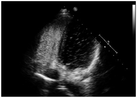
Fig. 2 Contrast transthoracic echocardiogram illustrating delayed right-to-left passage of agitated saline contrast (microbubbles) in left atrium and left ventricle.
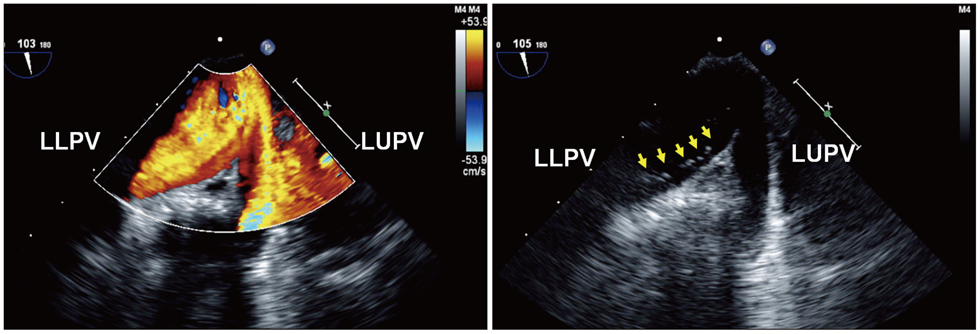
Fig. 3 Color flow imaging of transesophageal echocardiogram showing the inflow from left lower pulmonary vein (LLPV) and left upper pulmonary vein (LUPV). Contrast echocardiogram showing microbubbles in a row (arrows) draining into left atrium from LLPV but not from LUPV.
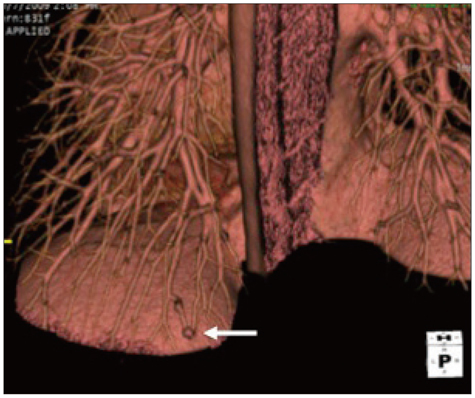
Fig. 4 Chest computed tomography angiogram with volume rendered reconstruction showing the fistula (arrow) located posteriorly in the left lower pulmonary lobe.
Reference
-
1. Ohara T, Nakatani S, Hashimoto S, Akaiwa Y, Yazaki S, Kimura K, Nakasone I, Masuda Y, Kanzaki H, Kitakaze M. A case of platypnea-orthodeoxia syndrome in a patient with a pulmonary arteriovenous fistula and a patent foramen ovale. J Am Soc Echocardiogr. 2007. 20:439.e5–439.e10.
Article
- Full Text Links
-
- Actions
-
Cited
- CITED
-
- Close
- Share
-
- Similar articles
-
- A Case Report of Coronary Arteriovenous Fistula Diagnosed by Two-Dimensional and Transesophageal Echocardiography
- A Case of Coronary Arteriovenous Fistula Confirmed by Echocardiography
- Arterio-Venous Line Connection for Effective Intracardiac Deairing after Open Heart Surgery
- A Case of Coronary Arteriovenous Fistula Associated with Pulmonary Artery Aneurysm Confirmed by Multi Detector-Row Helical CT
- A Case of Percutaneous Transcatheter Coil Embolization for Congenital Coronary Arteriovenous Fistula
