Transcatheter Treatment of Patent Foramen Ovale Combined with Abnormal Drainage of Left Superior Vena Cava to Left Upper Pulmonary Vein
- Affiliations
-
- 1Division of Pediatric Cardiology, Severance Cardiovascular Hospital, Yonsei University Health System, Seoul, Korea. cjy0122@yuhs.ac
- KMID: 1428376
- DOI: http://doi.org/10.4250/jcu.2013.21.1.33
Abstract
- Patent foramen ovale (PFO) has been known to be the cause of transient ischemic attacks or stroke, and transcatheter device closure has been the treatment of choice for these defects. Combined defect of abnormal drainage of left superior vena cava (LSVC) to left superior pulmonary vein (LSPV) in PFO patients is an uncommon combination, and both can act as a pathway for paradoxical embolism. We report a successful closure of PFO, using Amplatzer(R) PFO occluder (St. Jude Medical, St. Paul, MN, USA) and persistent LSVC connected to LSPV using an Amplatzer(R) Vascular Plug II (St. Jude Medical, St. Paul, MN, USA). Because this combined anomaly of PFO and persistent LSVC can be treated by a single transcatheter intervention, if clinically suspected, a complete evaluation for this anomaly should be considered.
Keyword
MeSH Terms
Figure
-
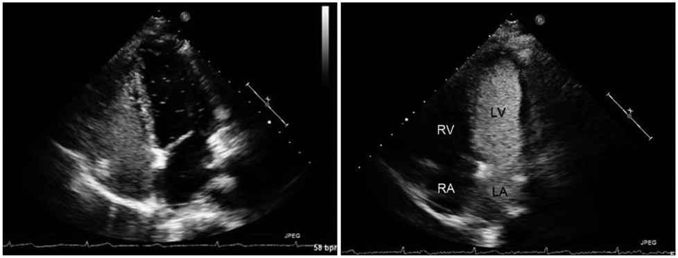
Fig. 1 Agitation saline injection test. On contrast echo conducted on Rt. arm, the enhancement was seen at not only Rt. side of heart, but also Lt. side. Sequential filling of the bubble in the left atrium followed by left ventricle and then through the patent foramen ovale to the right atrium. On Lt. arm contrast echocardiography, massive amount of microbubbles draining straight to LA was detected. LA: left atrium, LV: left ventricle, RA: right atrium, RV: right ventricle.
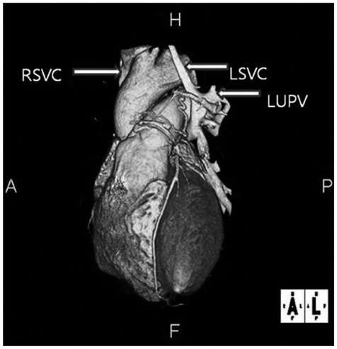
Fig. 2 Abnormal Drainage of LSVC to LUPV are shown on three dimensional computed tomography. LSVC: left superior vena cava, LUPV: left upper pulmonary vein, RSVC: right superior vena cava.
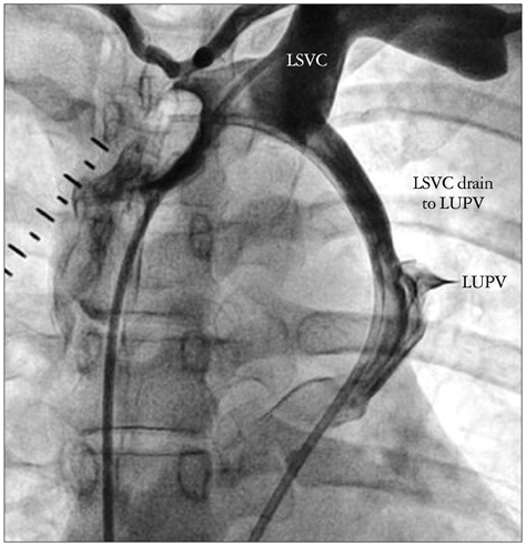
Fig. 3 Angiogram. Abnormal connection of LSVC to left atrium was shown on angiogram. LSVC: left superior vena cava, LUPV: left upper pulmonary vein.

Fig. 4 Abnormal drainage of left superior vena cava and left upper pulmonary vein was closed using the Amplatzer® vascular plug II.

Fig. 5 Transcatheter PFO closure with Amplatzer® PFO occluder. PFO: patent foramen ovale.
Reference
-
1. Irwin B, Ray S. Patent foramen ovale--assessment and treatment. Cardiovasc Ther. 2012. 30:e128–e135.
Article2. Tobis J, Shenoda M. Percutaneous treatment of patent foramen ovale and atrial septal defects. J Am Coll Cardiol. 2012. 60:1722–1732.
Article3. Biffi M, Boriani G, Frabetti L, Bronzetti G, Branzi A. Left superior vena cava persistence in patients undergoing pacemaker or cardioverter-defibrillator implantation: a 10-year experience. Chest. 2001. 120:139–144.
Article4. Winter FS. Persistent left superior vena cava; survey of world literature and report of thirty additional cases. Angiology. 1954. 5:90–132.5. Meadows WR, Sharp JT. Persistent left superior vena cava draining into the left atrium without arterial oxygen unsaturation. Am J Cardiol. 1965. 16:273–279.
Article6. Steinberg I, Dubilier W Jr, Lukas DS. Persistence of left superior vena cava. Dis Chest. 1953. 24:479–488.7. Fraser RS, Dvorkin J, Rossall RE, Eidem R. Left superior vena cava: a review of associated congenital heart lesions, catheterization data and roentgenologic findings. Am J Med. 1961. 31:711–716.8. Troost E, Gewillig M, Budts W. Percutaneous closure of a persistent left superior vena cava connected to the left atrium. Int J Cardiol. 2006. 106:365–366.
Article9. Wang W, Li H, Tam MD, Zhou D, Wang DX, Spain J. The amplatzer vascular plug: a review of the device and its clinical applications. Cardiovasc Intervent Radiol. 2012. 35:725–740.
Article10. Hutyra M, Skala T, Sanak D, Novotny J, Köcher M, Taborsky M. Persistent left superior vena cava connected through the left upper pulmonary vein to the left atrium: an unusual pathway for paradoxical embolization and a rare cause of recurrent transient ischaemic attack. Eur J Echocardiogr. 2010. 11:E35.
Article
- Full Text Links
-
- Actions
-
Cited
- CITED
-
- Close
- Share
-
- Similar articles
-
- Paradoxical Cerebral Embolism in a Young Patient: A case report
- A Case of Ischemic Colitis with Deep Vein Thrombosis and Patent Foramen Ovale
- Congenital Absence of the Azygos Vein with Persistent Left Superior Vena Cava: A Case Report
- Transcatheter Closure of Patent Foramen Ovale in a Stroke Patient under the Guidance of Transesophageal Echocardiography
- A Case of Persistent Left Superior Vena Cava with Interruption of Inferior Vena Cava
