J Korean Neurotraumatol Soc.
2009 Dec;5(2):115-117. 10.13004/jknts.2009.5.2.115.
An Unusual Cervical Post-Traumatic Pseudomeningocele: A Case Report
- Affiliations
-
- 1Department of Neurosurgery, Ilsan Paik Hospital, Inje University College of Medicine, Goyang, Korea. hsrkmj@paik.ac.kr
- KMID: 1427370
- DOI: http://doi.org/10.13004/jknts.2009.5.2.115
Abstract
- Pseudomeningoceles are formed by extravasation of cerebrospinal fluid through a dural defect into soft tissue. Most pseudomeningoceles are iatrogenic and occur in the posterior lumbar region following surgery. But, post-traumatic pseudomeningocele rarely occurs in the head and neck. We present a case of post-traumatic an unusual cervical pseudomenigocele and review the related literatures.
Keyword
MeSH Terms
Figure
-

FIGURE 1 Cervical enhanced CT scans (A: axial, B: coronal, C: sagittal) showed a massive fluid collection in the right anterior cervical region, which revealed a huge-sized pseudomeningocele arising from the right C6-7 intervertebral foramen (arrow).
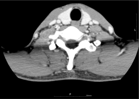
FIGURE 2 Follow-up cervical CT scan showed resolution of the pseudomeningocele.
Reference
-
1. Andrew SA, Sidhu KS. Cervical-peritoneal shunt placement for postoperative cervical pseudomeningocele. J Spinal Disord Tech. 2005; 18:290–292.2. Hawk MW, Kim KD. Review of spinal pseudomeningoceles and cerebrospinal fluid fistulas. Neurosurg Focus. 2000; 9:e5.
Article3. Horn EM, Bristol RE, Feiz-Erfan I, Beres EJ, Bambakidis NC, Theodore N. Spinal cord compression from traumatic anterior cervical pseudomeningoceles. Report of three cases. J Neurosurg Spine. 2006; 5:254–258.4. Hosono N, Yonenobu K, Ono K. Postoperative cervical pseudomeningocele with herniation of the spinal cord. Spine (Phila Pa 1976). 1995; 20:2147–2150.
Article5. Jeong JH, Ahn SK, Jeon SY, Park JJ, Kim JP, Park IS. Post-traumatic pseudomeningocele presenting as a cyst of external auditory canal: report of a case. Auris Nasus Larynx. 2006; 33:321–324.
Article6. Kitchen N, Bradford R, Platts A. Occult spinal pseudomeningocele following a trivial injury successfully treated with a lumboperitoneal shunt: a case report. Surg Neurol. 1992; 38:46–49.
Article7. Lau KK, Stebnyckyj M, McKenzie A. Post-laminectomy pseudomeningocele: an unusual cause of bone erosion. Australas Radiol. 1992; 36:262–264.
Article8. Maycock NF, van Essen J, Pfitzner J. Post-laminectomy cerebrospinal fluid fistula treated with epidural blood patch. Spine (Phila Pa 1976). 1994; 19:2223–2225.
Article9. McCormack BM, Taylor SL, Heath S, Scanlon J. Pseudomeningocele/CSF fistula in a patient with lumbar spinal implants treated with epidural blood patch and a brief course of closed subarachnoid drainage. A case report. Spine (Phila Pa 1976). 1996; 21:2273–2276.10. Naso WB, Cure J, Cuddy BG. Retropharyngeal pseudomeningocele after atlanto-occipital dislocation: report of two cases. Neurosurgery. 1997; 40:1288–1290.
Article11. Rinaldi I, Hodges TO. Iatrogenic lumbar meningocoele: report of three cases. J Neurol Neurosurg Psychiatry. 1970; 33:484–492.
Article12. Ryall RG, Peacock MK, Simpson DA. Usefulness of beta 2-transferrin assay in the detection of cerebrospinal fluid leaks following head injury. J Neurosurg. 1992; 77:737–739.13. Shapiro SA, Scully T. Closed continuous drainage of cerebrospinal fluid via a lumbar subarachnoid catheter for treatment or prevention of cranial/spinal cerebrospinal fluid fistula. Neurosurgery. 1992; 30:241–245.
Article14. Tate S, Rak RA, Bailey JS. Unusual presentation of a cervical pseudomeningocele: a case report and review of the literature. J Oral Maxillofac Surg. 2005; 63:556–559.
Article15. Taveras JM, Ransohoff J. Leptomeningeal cysts of the brain following trauma with erosion of the skull; a study of seven cases treated by surgery. J Neurosurg. 1953; 10:233–241.
- Full Text Links
-
- Actions
-
Cited
- CITED
-
- Close
- Share
-
- Similar articles
-
- A Case of Post-Traumatic Pseudomeningocele Treated by Lumboperitoneal Shunt
- A Case of Post-Traumatic Pseudomeningocele Presenting as a Cyst of External Auditory Canal
- Pseudomeningocele After Spine Surgery: 3 cases of different symptoms
- Pisiformectomy in Post-traumatic Pisotriquetral Osteoarthritis: A Case Report
- Clinical Experience of Pain Management for Postlaminectomy Syndrome due to Pseudomeningocele: A case report
