J Korean Soc Surg Hand.
2012 Dec;17(4):200-205. 10.12790/jkssh.2012.17.4.200.
Triceps Tendon Avulsion with a Bony Flake from the Olecranon: Three Cases Report
- Affiliations
-
- 1Department of Orthopedic Surgery, Konyang University College of Medicine, Daejeon, Korea. hurym1973@hanmail.net
- KMID: 1425264
- DOI: http://doi.org/10.12790/jkssh.2012.17.4.200
Abstract
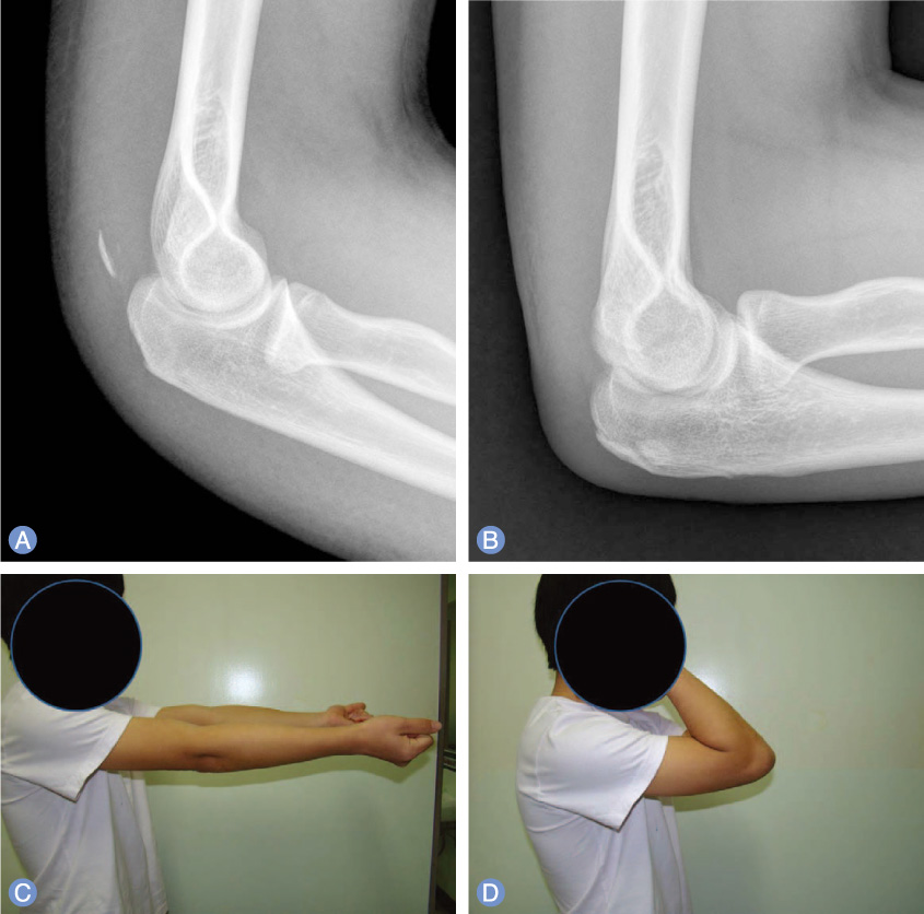
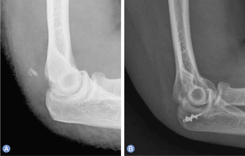
- Triceps tendon avulsion from the olecranon is a rare injury. Missed or delayed diagnosis may result in a weakness of strength or a decreased range of motion of the elbow. This injury is usually caused by a fall on the outstretched hand or a direct blow to the posterior arm. In addition, the rupture of the triceps tendon is implicated by a sudden eccentric contraction of the triceps muscle. To determine whether the rupture is complete or incomplete is critical to guide the treatment method. A small avulsed fragment from the olecranon may be detected on lateral radiographs of the elbow. We report three cases of the triceps tendon avulsion with a bony flake from the olecranon, which were surgically treated, along with a brief review of the literature.
Keyword
MeSH Terms
Figure
Reference
-
1. Anzel SH, Covey KW, Weiner AD, Lipscomb PR. Disruption of muscles and tendons; an analysis of 1, 014 cases. Surgery. 1959. 45:406–414.2. Mair SD, Isbell WM, Gill TJ, Schlegel TF, Hawkins RJ. Triceps tendon ruptures in professional football players. Am J Sports Med. 2004. 32:431–434.
Article3. Yeh PC, Dodds SD, Smart LR, Mazzocca AD, Sethi PM. Distal triceps rupture. J Am Acad Orthop Surg. 2010. 18:31–40.
Article4. van Riet RP, Morrey BF, Ho E, O'Driscoll SW. Surgical treatment of distal triceps ruptures. J Bone Joint Surg Am. 2003. 85:1961–1967.
Article5. Downey R, Jacobson JA, Fessell DP, Tran N, Morag Y, Kim SM. Sonography of partial-thickness tears of the distal triceps brachii tendon. J Ultrasound Med. 2011. 30:1351–1356.
Article6. Bach BR Jr, Warren RF, Wickiewicz TL. Triceps rupture. A case report and literature review. Am J Sports Med. 1987. 15:285–289.7. Lambers K, Ring D. Elbow fracture-dislocation with triceps avulsion: report of 2 cases. J Hand Surg Am. 2011. 36:625–627.
Article8. Farrar EL 3rd, Lippert FG 3rd. Avulsion of the triceps tendon. Clin Orthop Relat Res. 1981. (161):242–246.
Article9. Weng PW, Wang SJ, Wu SS. Misdiagnosed avulsion fracture of the triceps tendon from the olecranon insertion: case report. Clin J Sport Med. 2006. 16:364–365.
Article


