Multilobular Lacrimal Sac Diverticulum Presenting as a Lower Eyelid Mass
- Affiliations
-
- 1Department of Ophthalmology, Inje University Sanggye Paik Hospital, Seoul, Korea.
- 2Department of Ophthalmology, Kangbuk Samsung Hospital, Sungkyunkwan University School of Medicine, Seoul, Korea.
- 3Department of Ophthalmology, Samsung Medical Center, Sungkyunkwan University School of Medicine, Seoul, Korea. eyeminded@skku.edu
- KMID: 1387398
- DOI: http://doi.org/10.3341/kjo.2012.26.4.297
Abstract
- Lacrimal sac diverticulum is a rare condition, and its various symptoms complicate differential diagnosis. We present cases of a peculiar type of lacrimal diverticulum. A 5-year-old girl and a 50-year-old woman presented with a protruding mass inferior to the medial canthus. Each lacrimal system was patent to irrigation. The masses compressed and distorted the lacrimal passage and had no apparent connection with the lacrimal sac in dacryocystography or computed tomography. Surgical exploration and complete excision of the masses were completed. Each patient had an inverted Y- and an inverted V-shaped multilobular cystic mass that was pathologically confirmed as a lacrimal sac diverticulum. Lacrimal sac diverticula may rarely take the form of a multilobular cyst and can present as a lower lid mass. We speculate that an abnormality in lacrimal embryogenesis resulted in multiple blind pouches, a peculiar type of lacrimal sac diverticulum.
Keyword
MeSH Terms
Figure
-
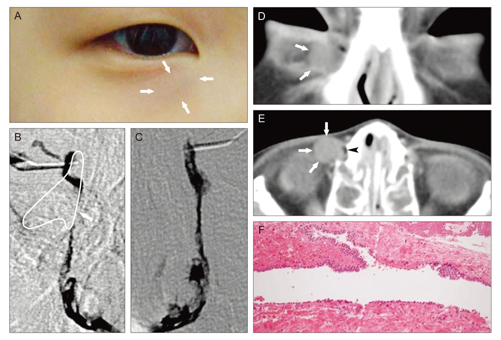
Fig. 1 Case 1. (A) A mass was found along the right inferior orbital rim (arrows). (B) Dacryocystography was performed. A reconstructed image of an inverted Y-shaped mass (white line): the lateral lobule extended along the inferior orbital rim, the inferior lobule pushed the nasolacrimal duct medially (arrow), and the superior lobule displaced the lacrimal sac both anteriorly and laterally. There was no dye outflow across the lacrimal sac wall. (C) Dacryocystography of the left side. (D) Coronal computed tomography (CT): a soft tissue mass surrounded by rim enhancement (arrows). (E) Axial CT: air density (arrowhead) of the lacrimal sac was displaced anteromedially by the mass (arrows). The lacrimal sac fossa was widened. (F) Epithelium of the mass consisted of pseudo-stratified cuboidal and columnar epithelium. The wall of the mass consisted of fibroblasts and collagen fibers (H&E, ×100).
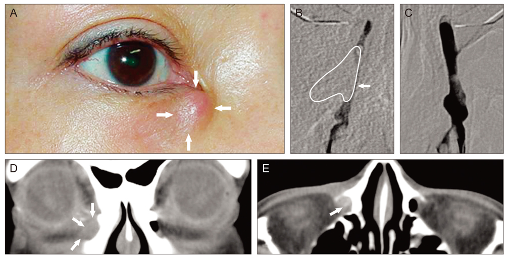
Fig. 2 Case 2. (A) A lower eyelid mass fixed along the inferior orbital rim (arrows) with overlying skin erythema. (B) Dacryocystograph of the right side: a reconstructed image of an inverted V-shaped mass (white line) next to the right lacrimal sac and nasolacrimal duct. The mass was compressing the lacrimal sac and nasolacrimal duct (arrow). (C) Dacryocystograph of the left side. (D) Coronal computed tomography (CT): a soft tissue mass (arrows) compressed the lacrimal sac at the level of the medial canthus. (E) Axial CT: a high density inside the mass (arrow) at the level of the upper part of the nasolacrimal duct indicated dacryolithiasis at the inferior lobule of the diverticulum.
Reference
-
1. Duke-Elder S, MacFaul PA. Duke-Elder S, editor. The ocular adnexa: lacrimal, orbital, and para-orbital diseases. Cysts and diverticula. System of ophthalmology. Vol. 13. The ocular adnexa. 1974. St. Louis: CV Mosby;733–735.2. Polito E, Leccisotti A, Menicacci F, et al. Imaging techniques in the diagnosis of lacrimal sac diverticulum. Ophthalmologica. 1995. 209:228–232.3. Ormrod JN. Diverticulum of the lacrimal sac. Br J Ophthalmol. 1958. 42:526–528.4. Sinnreich Z. Lacrimal diverticula. Orbit. 1998. 17:195–200.5. Akcay EK, Cagil N, Yulek F, et al. Congenital lacrimal sac diverticulum as a cause of recurrent orbital cellulitis. Can J Ophthalmol. 2009. 44:e29–e30.6. Hurwitz JJ. Hurwitz JJ, editor. Embryology of the lacrimal drainage system. The lacrimal system. 1996. Philadelphia: Lippincott-Raven;9–13.7. Satchi K, McNab AA. Double lacrimal puncta: clinical presentation and potential mechanisms of epiphora. Ophthalmology. 2010. 117:180–183.e2.8. Hosal BM, Hurwitz JJ, Howarth DJ. Orbital cyst of lacrimal sac derivation. Eur J Ophthalmol. 1996. 6:279–283.
- Full Text Links
-
- Actions
-
Cited
- CITED
-
- Close
- Share
-
- Similar articles
-
- A Case of Congenital Lacrimal Sac Diverticulum
- A Case of Benign Mixed Tumor Presenting as a Nodular Eyelid Lesion
- Apocrine Hidrocystoma Presenting as a Lacrimal Gland Mass
- A Case of Primary Neurofibroma of Lacrimal Sac
- Small Cell Neuroendocrine Carcinoma of the Lacrimal Sac Presenting with Lacrimal Duct Obstruction
