Giant Cell Tumor of Soft Tissue: a Case with Atypical US and MRI Findings
- Affiliations
-
- 1Department of Radiology and Institute of Radiation Medicine, Seoul National University College of Medicine, Seoul, Korea. jacrad@radiol.snu.ac.kr
- 2Department of Radiology, Seoul National University Bundang Hospital, Gyeongi-do, Korea.
- 3Department of Pathology, Seoul National University Bundang Hospital, Gyeongi-do, Korea.
- 4Department of Orthopedic Surgery, Seoul National University Bundang Hospital, Gyeongi-do, Korea.
- KMID: 1385406
- DOI: http://doi.org/10.3348/kjr.2008.9.5.462
Abstract
- We report the case of a giant cell tumor with diffuse interstitial hemorrhaging and unusually prominent cystic components in the soft tissue of the thigh which has not been reported previously. Magnetic resonance image (MRI), showed signal intensity typical of a giant cell tumor. However, because of its conspicuous large well-circumscribed cystic components, the differential diagnoses, based on the image findings from an ultrasonography (US) and MRI, were complicated epidermoid cyst, cystic change of a neurogenic tumor, and a parasitic cyst.
Keyword
MeSH Terms
Figure
-
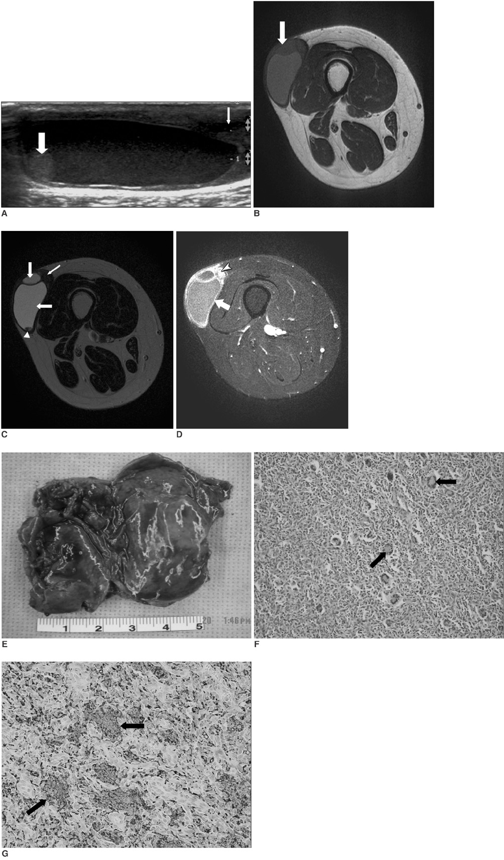
Fig. 1 23-year-old woman with giant cell tumor of soft tissue in thigh. A. Sonogram of lateral aspect of right thigh shows primary cyst with debris and hyperechoic nodule (thick arrow). Daughter cyst was located adjacent to main cyst (thin arrow) and is also seen with fluid-fluid levels within. B. Axial T1-weighted image (TR/TE, 430/20) shows cystic mass with intermediate signal intensity (arrow) adjacent to vastus lateralis muscle in subcutaneous tissue of right thigh. C. Axial T2-weighted image (TR/TE, 4039/100) shows cystic mass with low signal intensity, which comprised of two cysts (thick arrows) and solid portion (thin arrow). Note difference in signal intensity between two cysts. Nodule (arrowhead) within large cyst is well visualized. D. Contrast-enhanced T1-weighted image (TR/TE, 500/15) with fat suppression shows diffuse enhancement of cystic wall (arrow) and solid portion (arrowhead) of mass. E. Gross photograph of excised specimen revealing extensive cystic change of mass with dark-brown color. F. Histologic features of giant cell tumor showing cystic change. Cystic wall of this mass shows mixture of mononuclear spindle cells and multinucleated osteoclast-like giant cells (arrows, Hematoxylin & Eosin staining, ×100). G. Immunohistochemical staining for CD68. Tumor cells show diffuse positive reaction in multinucleated giant cells (arrows) and focally in mononuclear spindle cells (×400) for CD68.
Reference
-
1. Holst VA, Elenitsas R. Primary giant cell tumor of soft tissue. J Cutan Pathol. 2001. 28:492–495.2. Oliveira AM, Dei Tos AP, Fletcher CD, Nascimento AG. Primary giant cell tumor of soft tissues: a study of 22 cases. Am J Surg Pathol. 2000. 24:248–256.3. O'Connell JX, Wehrli BM, Nielsen GP, Rosenberg AE. Giant cell tumors of soft tissue: a clinicopathologic study of 18 benign and malignant tumors. Am J Surg Pathol. 2000. 24:386–395.4. Murphey MD, Nomikos GC, Flemming DJ, Gannon FH, Temple HT, Kransdorf MJ. From the crchives of AFIP. Imaging of giant cell tumor and giant cell reparative granuloma of bone: radiologic-pathologic correlation. Radiographics. 2001. 21:1283–1309.5. Aoki J, Moriya K, Yamashita K, Fujioka F, Ishii K, Karakida O, et al. Giant cell tumors of bone containing large amounts of hemosiderin: MR-pathologic correlation. J Comput Assist Tomogr. 1991. 15:1024–1027.6. Kransdorf MJ, Sweet DE. Aneurysmal bone cyst: concept, controversy, clinical presentation, and imaging. AJR Am J Roentgenol. 1995. 164:573–580.7. Tsai JC, Dalinka MK, Fallon MD, Zlatkin MB, Kressel HY. Fluid-fluid level: a nonspecific finding in tumors of bone and soft tissue. Radiology. 1990. 175:779–782.8. Lee EY, Kang KS, Kang SY, Lee HJ, Kim JW, Lee GH, et al. Soft tissue Giant Cell Tumor of Low Malignant Potential. J Korean Bone & Joint Tumor Soc. 2003. 9:101–104. (Korean).9. Dodd LG, Major N, Brigman B. Malignant giant cell tumor of soft parts. Skeletal Radiol. 2004. 33:295–299.
- Full Text Links
-
- Actions
-
Cited
- CITED
-
- Close
- Share
-
- Similar articles
-
- Ossified Soft Tissue Recurrence of Giant Cell Tumor: Three Case Report
- A Case of Soft Tissue Recurrence after Wide Resection of Giant Cell Tumor in the Distal Femur
- A Metastatic Giant Cell Tumor of the Soft Tissue of the Thoracic Wall: A case report
- MR Findings of Recurred Giant Cell Tumor
- MR Findings of Giant Cell Tumor: Signal Intensity and Morphological Characteristics
