Conventional Galactography and MR Contrast Galactography for Diagnosing Nipple Discharge: Preliminary Results
- Affiliations
-
- 1Department of Radiology, S.B. Ankara Diskapi Training and Research Hospital, Ankara, Turkey. yucecun2000@yahoo.com
- 2Department of General Surgery, S.B. Ankara Diskapi Training and Research Hospital, Ankara, Turkey.
- KMID: 1385401
- DOI: http://doi.org/10.3348/kjr.2008.9.5.426
Abstract
OBJECTIVE
We wanted to compare the clinical usefulness of conventional galactography and MR contrast galactography for diagnosing patients with nipple discharge. MATERIALS AND METHODS: Both conventional galactography and MR contrast galactography were performed prospectively in 16 patients. Gadopentate dimeglumine (0.1 ml) was mixed with non-ionic contrast medium (0.9 ml) to obtain a resultant volume of 1 ml and this was used for both examinations. Following conventional galactography, MR contrast galactography was performed after direct injection of contrast media into the duct. RESULTS: Conventional galactography and MR contrast galactography were concordant in 13 (81%) of 16 patients; the results were normal in five, ductal dilatation was noted in four and intraductal filling defects were noted in four. The remaining three (19%) patients demonstrated discordant findings on the two examinations. While conventional galactography revealed filling defects, the MR contrast galactography results were normal in two patients. The third patient had kinks-stricture on conventional galactography and MR contrast galactography showed ductal dilatation. This suggested there were false positive results for the three patients' conventional galactography, and all the three patients with discordant results underwent surgery and the histopathologic evaluation showed fibrocystic changes. CONCLUSION: MR contrast galactography may be used as an alternative imaging modality for making the diagnosis of pathologic nipple discharge. However, statistically supported studies with large pools of subjects for comparing the galactography and MR contrast galactography results are needed to confirm our findings.
MeSH Terms
Figure
-
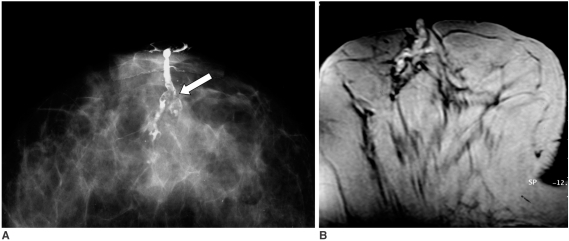
Fig. 1 49-year-old woman with bloody discharge.A. Conventional galactography on craniocaudal projection shows irregular filling defects (arrow). Note duct is extended due to compression.B. MR contrast galactography on axial plane at same level shows no filling defects and pathology revealed fibrocyctic changes. Note duct is minimally heterogeneous, possibly as result of different degrees of hemorrhage mixed with contrast material.

Fig. 2 54-year-old woman with bloody discharge.A. Conventional galactography on craniocaudal projection demonstrates dilated ducts and filling defects (arrows).B. MR contrast galactography on axial plane shows hyperintense dilated ducts and hypointense multiple filling defects (arrows). Pathology revealed intraductal papillomas.

Fig. 3 33-year-old woman with bloody discharge.A, B. Both conventional galactography (A) and MR contrast galactography (B) reveal filling defects (arrows) and pathology results were reported as intraductal papillomas.
Cited by 1 articles
-
Does Ultrasound-Guided Directional Vacuum-Assisted Removal Help Eliminate Abnormal Nipple Discharge in Patients with Benign Intraductal Single Mass?
Jung Min Chang, Nariya Cho, Woo Kyung Moon, Jeong Seon Park, Se-Yeong Chung, Mijung Jang
Korean J Radiol. 2009;10(6):575-580. doi: 10.3348/kjr.2009.10.6.575.
Reference
-
1. Dinkel HP, Trusen A, Gassel AM, Rominger M, Lourens S, Muller T, et al. Predictive value of galactographic patterns for benign and malignant neoplasms of the breast in patients with nipple discharge. Br J Radiol. 2000; 73:706–714. PMID: 11089460.
Article2. Cho N, Moon WK, Chung SY, Cha JH, Cho KS, Kim EK, et al. Ductographic findings of breast cancer. Korean J Radiol. 2005; 6:31–36. PMID: 15782017.
Article3. Funovics MA, Philipp MO, Lackner B, Fuchsjaeger M, Funovics PT, Metz V. Galactography: method of choice in pathologic nipple discharge? Eur Radiol. 2003; 13:94–99. PMID: 12541115.
Article4. Slawson SH, Johnson BA. Ductography: how to and what if? Radiographics. 2001; 21:133–150. PMID: 11158649.
Article5. Heywang Kobrunner SH. Contrast-enhanced magnetic resonance imaging of the breast. Invest Radiol. 1994; 29:94–104. PMID: 8144345.6. Harms SE, Flamig DP. MR imaging of the breast. J Magn Reson Imaging. 1993; 3:277–283. PMID: 8428095.
Article7. Gilles R, Guinebretiere JM, Lucidarme O, Cluzel P, Janaud G, Finet JF, et al. Nonpalpable breast tumors: diagnosis with contrast-enhanced subtraction dynamic MR imaging. Radiology. 1994; 191:625–631. PMID: 8184038.
Article8. Sardanelli F, Podo F, D'Agnolo G, Verdecchia A, Santaquilani M, Musumeci R, et al. Multicenter comparative multimodality surveillance of women at genetic-familial high risk for breast cancer (HIBCRIT Study): Interim Results. Radiology. 2007; 242:698–715. PMID: 17244718.
Article9. Kim do Y, Moon WK, Cho N, Ko ES, Yang SK, Park JS, et al. MRI of the breast for the detection and assessment of the size of ductal carcinoma in situ. Korean J Radiol. 2007; 8:32–39. PMID: 17277561.
Article10. American College of Radiology. Breast imaging reporting and data system (BIRADS). 2003. Reston, VA: American College of Radiology.11. Baker KS, Davey DD, Stelling CB. Ductal abnormalities detected with galactography: frequency of adequate excisional biopsy. AJR Am J Roentgenol. 1994; 162:821–824. PMID: 8140998.
Article12. Cardenosa G, Doudna C, Eklund GW. Ductography of the breast: technique and findings. AJR Am J Roentgenol. 1994; 162:1081–1087. PMID: 8165986.
Article13. Orel SG, Dougherty CS, Reynolds C, Czerniecki BJ, Siegelman ES, Schnall MD. MR imaging in patients with nipple discharge: initial experience. Radiology. 2000; 216:248–254. PMID: 10887256.
Article14. Rovno HD, Sigelman ES, Reynolds C, Orel SG, Schnall MD. Solitary intraductal papilloma: findings at MR imaging and MR galactography. AJR Am J Roentgenol. 1999; 172:151–155. PMID: 9888758.
Article15. Yoshimoto M, Kasumi F, Iwase T, Takahashi K, Tada T, Uchida Y. Magnetic resonance galactography for a patient with nipple discharge. Breast Cancer Res Treat. 1997; 42:87–90. PMID: 9116323.
Article16. Kanemaki Y, Kurihara Y, Itoh D, Kamijo K, Nakajima Y, Fukuda M, et al. MR mammary ductography using a microscopy coil for assessment of intraductal lesions. AJR Am J Roentgenol. 2004; 182:1340–1342. PMID: 15100142.
Article17. Bhattarai N, Kanemaki Y, Kurihara Y, Nakajima Y, Fukuda M, Maeda I. Intraductal papilloma: features on MR ductography using a microscopic coil. AJR Am J Roentgenol. 2006; 186:44–47. PMID: 16357375.
Article
- Full Text Links
-
- Actions
-
Cited
- CITED
-
- Close
- Share
-
- Similar articles
-
- Breast tumors associated with nipple discharge: US findings with galactographic correlation
- Galactography in non-lactating nipple discharge
- Values of contrast galactography in nonlactating nipple discharge in Korean women
- Galactographic Differentiation between Malignant and Benign Disease in Patients with Pathologic Nipple Discharge
- Solitary Breast Papilloma: Comparison of Galactographic and Ductal
