Solitary Pulmonary Nodule on Helical Dynamic CT Scans: Analysis of the Enhancement Patterns Using a Computer-Aided Diagnosis (CAD) System
- Affiliations
-
- 1Department of Radiology, Chonbuk National University Hospital and Medical School, Research Institute for Medical Science, Chonbuk, Korea. gyjin@chonbuk.ac.kr
- 2Department of Province, Chonbuk National University Hospital and Medical School, Research Institute for Medical Science, Chonbuk, Korea.
- KMID: 1385397
- DOI: http://doi.org/10.3348/kjr.2008.9.5.401
Abstract
OBJECTIVE
We wanted to investigate the usefulness of a computer-aided diagnosis (CAD) system in assisting radiologists to diagnosis malignant solitary pulmonary nodules (SPNs), as compared with diagnosing SPNs with using direct personal drawing. MATERIALS AND METHODS: Forty patients with SPNs were analyzed. After the pre-contrast scan was performed, an additional ten series of post-contrast images were obtained at 20-second intervals. Two investigators measured the attenuation values of the SPNs: a radiologist who drew the regions of interest (ROIs), and a technician who used a CAD system. The Bland and Altman plots were used to compare the net enhancement between a CAD system and direct personal drawing. The diagnostic characteristics of the malignant SPNs were calculated by considering the CAD and direct personal drawing and with using Fisher's exact test. RESULTS: On the Bland and Altman plot, the net enhancement difference between the CAD system and direct personal drawing was not significant (within +/- 2 standard deriation). The sensitivity, specificity, positive predictive value (PPV), negative predictive value (NPV) and accuracy of diagnosing malignant SPNs using CAD was 92%, 85%, 75%, 96% and 88%, respectively. The sensitivity, specificity, PPV, NPV and accuracy of diagnosing malignant SPNs using direct drawing was 92%, 89%, 79%, 92% and 88%, respectively. CONCLUSION: The CAD system was a useful tool for diagnosing malignant SPNs.
MeSH Terms
-
Adult
Aged
Contrast Media
Diagnosis, Computer-Assisted/*methods
Diagnosis, Differential
Female
Humans
Iohexol/analogs & derivatives/diagnostic use
Lung Neoplasms/*radiography
Male
Middle Aged
Predictive Value of Tests
Radiographic Image Enhancement/*methods
Retrospective Studies
Sensitivity and Specificity
Solitary Pulmonary Nodule/*radiography
Tomography, Spiral Computed/*methods
Figure
-
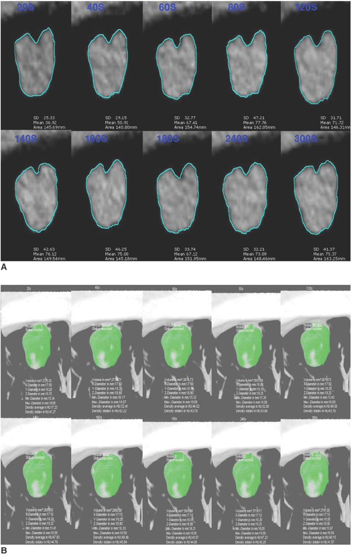
Fig. 1 Methods of enhancement pattern analysis using CAD system. A. Before intravenously injecting contrast medium, series of images was obtained throughout entire nodule along z-axis, and additional ten sets (20s, 40s, 60s, 80s, 120s, 140s, 160s, 180s, 240s and 300s) of images were obtained at 20-second intervals over 5-minute period after injecting contrast medium. Region of interest covered approximately full diameter of nodule and single radiologist directly drew region of interest. B. Solitary pulmonary nodule was region of interest drawn by expert technician with using CAD system.

Fig. 2 Bland and Altman plot showing difference between using CAD system and direct personal drawing. A. Difference in peak enhancement between using CAD system and direct personal drawing. B. Difference in net enhancement between using CAD system and direct personal drawing. X-axis represented average values of using CAD system and direct personal drawing while y-axis represented difference in peak enhancement and net enhancement between using CAD system and direct personal drawing. Dashed lines represent mean value of two measurements and lines above and below it represent 95% limits of agreement. Peak enhancement was out of 95% limits of agreement only in one case, while net enhancement was out of 95% limits of agreement in two cases.
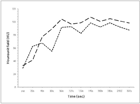
Fig. 3 CT scan of a benign solitary pulmonary nodule with enhancement (≥15 HU wash-in, ≥25 HU washout) in 43-year-old female diagnosed with sclerosing hemangioma by open lung biopsy. Time attenuation curve obtained through nodule for 5 minutes showed similar enhancement patterns of CAD system (short line) and direct personal drawing (long line).

Fig. 4 CT scan of adenocarcinoma with enhancement (≥15 HU wash-in, 5-25 HU washout) in 72-year-old man. Time attenuation curve obtained through nodule for 5 minutes showed similar enhancement patterns of CAD system (short line) and direct personal drawing (long line).
Reference
-
1. Jeong YJ, Yi CA, Lee KS. Solitary pulmonary nodules: detection, characterization, and guidance for further diagnostic workup and treatment. AJR Am J Roentgenol. 2007. 188:57–68.2. Leef JL 3rd, Klein JS. The solitary pulmonary nodule. Radiol Clin North Am. 2002. 40:123–143.3. Shaham D, Guralnik L. The solitary pulmonary nodule: radiologic considerations. Semin Ultrasound CT MR. 2000. 21:97–115.4. Tan BB, Flaherty KR, Kazerooni EA, Iannettoni MD. The solitary pulmonary nodule. Chest. 2003. 123:S89–S96.5. Tang AW, Moss HA, Robertson RJ. The solitary pulmonary nodule. Eur J Radiol. 2003. 45:69–77.6. Wang MP, Tan YQ, Zhang GZ, Zhang JG, Wu H, Yang JY. Differential diagnosis of benign and malignant solitary pulmonary nodule with computer-aided detection. Zhongguo Yi Xue Ke Xue Yuan Xue Bao. 2006. 28:64–67.7. Erasmus JJ, Connolly JE, McAdams HP, Roggli VL. Solitary pulmonary nodules: Part I. Morphologic evaluation for differentiation of benign and malignant lesions. Radiographics. 2000. 20:43–58.8. Marten K, Grabbe E. The challenge of the solitary pulmonary nodule: diagnostic assessment with multislice spiral CT. Clin Imaging. 2003. 27:156–161.9. Yankelevitz DF, Henschke CI. Small solitary pulmonary nodules. Radiol Clin North Am. 2000. 38:471–478.10. Yi CA, Lee KS, Kim BT, Choi JY, Kwon OJ, Kim H, et al. Tissue characterization of solitary pulmonary nodule: comparative study between helical dynamic CT and integrated PET/CT. J Nucl Med. 2006. 47:443–450.11. Trotman-Dickenson B, Baumert B. Multidetector-row CT of the solitary pulmonary nodule. Semin Roentgenol. 2003. 38:158–167.12. Das M, Mühlenbruch G, Mahnken AH, Flohr TG, Gündel L, Stanzel S, et al. Solitary pulmonary nodule: effect of two computer-aided detection systems on radiologist performance. Radiology. 2006. 241:564–571.13. Muhm JR, McCullough AE. The enhancing rim: a new sign of a benign pulmonary nodule. Mayo Clin Proc. 2003. 78:1092–1096.14. Swensen SJ, Viggiano RW, Midthun DE, Müller NL, Sherrick A, Yamashita K, et al. Lung nodule enhancement at CT: multicenter study. Radiology. 2000. 214:73–80.15. Jeong YJ, Lee KS, Jeong SY, Chung MJ, Shim SS, Kim H, et al. Solitary pulmonary nodule: characterization with combined wash-in and washout features at dynamic multi-detector row CT. Radiology. 2005. 237:675–683.16. Schaefer JF, Vollmar J, Schick F, Vonthein R, Seemann MD, Aebert H, et al. Solitary pulmonary nodules: dynamic contrast-enhanced MR imaging-perfusion differences in malignant and benign lesions. Radiology. 2004. 232:544–553.17. Yi CA, Lee KS, Kim EA, Han J, Kim H, Kwon OJ, et al. Solitary pulmonary nodules: dynamic enhanced multi-detector row CT study and comparison with vascular endothelial growth factor and microvessel density. Radiology. 2004. 233:191–199.18. Shah SK, McNitt-Gray MF, Rogers SR, Goldin JG, Suh RD, Sayre JW, et al. Computer aided characterization of the solitary pulmonary nodule using volumetric and contrast enhancement features. Acad Radiol. 2005. 12:1310–1319.19. Shah SK, McNitt-Gray MF, Rogers SR, Goldin JG, Suh RD, Sayre JW, et al. Computer-aided diagnosis of the solitary pulmonary nodule. Acad Radiol. 2005. 12:570–575.20. Mori K, Niki N, Kondo T, Kamiyama Y, Kodama T, Kawada Y, et al. Development of a novel computer-aided diagnosis system for automatic discrimination of malignant from benign solitary pulmonary nodules on thin-section dynamic computed tomography. J Comput Assist Tomogr. 2005. 29:215–222.21. Lee JW, Goo JM, Lee HJ, Kim JH, Kim S, Kim YT. The potential contribution of a computer-aided detection system for lung nodule detection in multidetector row computed tomography. Invest Radiol. 2004. 39:649–655.22. Kim KG, Goo JM, Kim JH, Lee HJ, Min BG, Bae KT, et al. Computer-aided diagnosis of localized ground-glass opacity in the lung at CT: initial experience. Radiology. 2005. 237:657–661.23. Goo JM, Lee JW, Lee HJ, Kim S, Kim JH, Im JG. Automated lung nodule detection at low-dose CT: preliminary experience. Korean J Radiol. 2003. 4:211–216.24. Rubin GD, Lyo JK, Paik DS, Sherbondy AJ, Chow LC, Leung AN, et al. Pulmonary nodules on multi-detector row CT scans: performance comparison of radiologists and computer-aided detection. Radiology. 2005. 234:274–283.25. Verdun FR, Gutierrez D, Schnyder P, Aroua A, Bochud F, Gudinchet F. CT dose optimization when changing to CT multi-detector row technology. Curr Probl Diagn Radiol. 2007. 36:176–184.26. Bland JM, Altman DG. Statistics methods of assessing agreement between two methods of clinical measurement. Lancet. 1986. 1:307–310.27. McNitt-Gray MF. AAPM/RSNA physics tutorial for residents: topics in CT. Radiation dose in CT. Radiographics. 2002. 22:1541–1553.
- Full Text Links
-
- Actions
-
Cited
- CITED
-
- Close
- Share
-
- Similar articles
-
- A Computer-Aided Diagnosis for Evaluating Lung Nodules on Chest CT: the Current Status and Perspective
- Computer-Aided Differential Diagnosis of the Pulmonary Nodule: Towards an Understanding of the Medical Imaging Basics and Experiences in the Field
- Studies and Real-World Experience Regarding the Clinical Application of Artificial Intelligence Software for Lung Nodule Detection
- Malignant Solitary Pulmonary Nodule: Enhancement Patterns on Contrast-enhanced Dynamic CT with the Histopathologic Evaluation
- The Role of Dynamic CT for the Differential Diagnosis of Solitary Pulmonary Nodule
