Spectral CT: Preliminary Studies in the Liver Cirrhosis
- Affiliations
-
- 1Department of Radiology, The First Affiliated Hospital of Zhengzhou University, Henan Province 450052, China.
- 2Department of Radiology, Ruijin Hospital, Shanghai Jiao Tong University School of Medicine, Shanghai 200025, China. keminchen0307@yahoo.com.cn
- KMID: 1383855
- DOI: http://doi.org/10.3348/kjr.2012.13.4.434
Abstract
OBJECTIVE
To investigate the value of spectral CT imaging in the diagnosis and classification of liver cirrhosis during the arterial phase (AP) and portal venous phase (PVP).
MATERIALS AND METHODS
Thirty-eight patients with liver cirrhosis (Child-Pugh class A/B/C: n = 10/14/14), and 43 patients with healthy livers, participated in this study. The researchers used abdominal spectral CT imaging during AP and PVP. Iodine concentration, derived from the iodine-based material-decomposition image and the iodine concentration ratio (ICratio) between AP and PVP, were obtained. Statistical analyses {two-sample t test, One-factor analysis of variance, and area under the receiver operating characteristic curve (A [z])} were performed.
RESULTS
The mean normalized iodine concentration (NIC) (0.5 +/- 0.12) during PVP in the control group was significantly higher than that in the study group (0.4 +/- 0.10 on average, 0.4 +/- 0.08 for Class A, 0.4 +/- 0.15 for Class B, and 0.4 +/- 0.06 for Class C) (All p < 0.05). Within the cirrhotic liver group, the mean NIC for Class C during the AP (0.1 +/- 0.05) was significantly higher than NICs for Classes A (0.1 +/- 0.06) and B (0.1 +/- 0.03) (Both p < 0.05). The ICratio in the study group (0.4 +/- 0.15), especially for Class C (0.5 +/- 0.14), was higher than that in the control group (0.3 +/- 0.15) (p < 0.05).The combination of NIC and ICratio showed high sensitivity and specificity for differentiating healthy liver from cirrhotic liver, especially in Class C cirrhotic liver.
CONCLUSION
Spectral CT Provides a quantitative method with which to analyze the cirrhotic liver, and shows the potential value in the classification of liver cirrhosis.
MeSH Terms
Figure
-
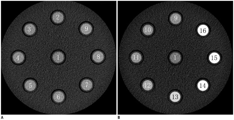
Fig. 1 Distribution of 16 tubes with different concentrations of iodine solution.Arrangement of 16 tubes (A, B) containing iodine solutions with different concentrations (0 to 30.0 mg/mL) at transverse monochromatic CT image obtained at 65-keV energy level.
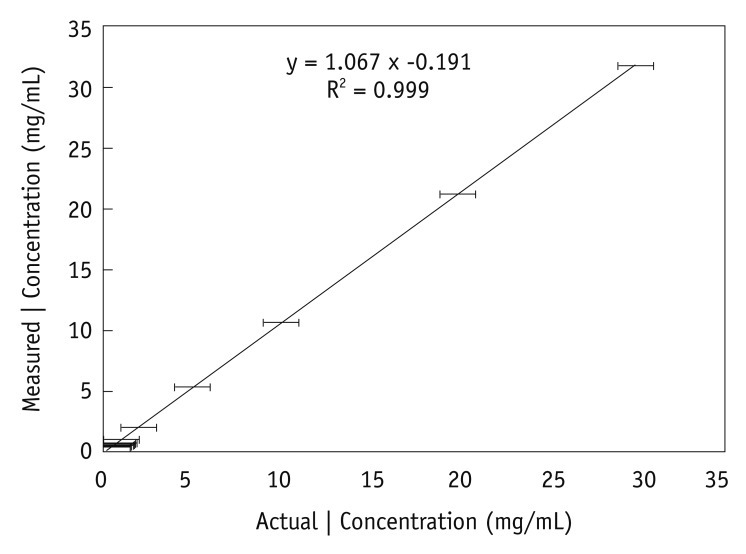
Fig. 2 Graph illustrates relationship between actual (x-axis) and measured (y-axis) iodine (I) concentrations in in vitro experiment. Test tubes filled with known iodine concentrations of 0.3-30.0 mg/mm3 were scanned with CT spectral imaging, and iodine concentrations were measured from iodine-based material-decomposition images. Mean and standard deviations for three separate measurements for same iodine concentration in each test tube (n = 13) are shown. Fitted line shows linear relationship between measured and actual concentrations.
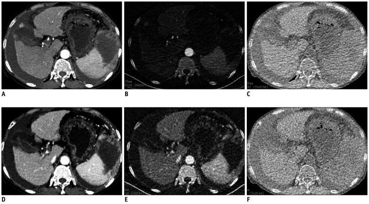
Fig. 3 Monochromatic and material decomposition images of cirrhosis.Transverse (A, D) monochromatic CT image obtained at 70-keV energy level and (B, E) iodine-based and (C, F) water-based material-decomposition images obtained from single spectral CT acquisition (section thickness, 1.25 mm) in 63-year-old woman with cirrhosis B during AP and PVP respectively. AP = arterial phase, PVP = portal venous phase
Reference
-
1. Genant HK, Boyd D. Quantitative bone mineral analysis using dual energy computed tomography. Invest Radiol. 1977; 12:545–551. PMID: 591258.
Article2. Chiro GD, Brooks RA, Kessler RM, Johnston GS, Jones AE, Herdt JR, et al. Tissue signatures with dual-energy computed tomography. Radiology. 1979; 131:521–523. PMID: 441344.
Article3. Millner MR, McDavid WD, Waggener RG, Dennis MJ, Payne WH, Sank VJ. Extraction of information from CT scans at different energies. Med Phys. 1979; 6:70–71. PMID: 440238.
Article4. Louis O, Van den Winkel P, Covens P, Schoutens A, Osteaux M. Mineral content of vertebral trabecular bone: accuracy of dual energy quantitative computed tomography evaluated against neutron activation analysis and flame atomic absorption spectrometry. Bone. 1994; 15:35–39. PMID: 8024849.
Article5. Kelcz F, Joseph PM, Hilal SK. Noise considerations in dual energy CT scanning. Med Phys. 1979; 6:418–425. PMID: 492076.
Article6. Coursey CA, Nelson RC, Boll DT, Paulson EK, Ho LM, Neville AM, et al. Dual-energy multidetector CT: how does it work, what can it tell us, and when can we use it in abdominopelvic imaging? Radiographics. 2010; 30:1037–1055. PMID: 20631367.
Article7. Lv P, Lin XZ, Li J, Li W, Chen K. Differentiation of small hepatic hemangioma from small hepatocellular carcinoma: recently introduced spectral CT method. Radiology. 2011; 259:720–729. PMID: 21357524.
Article8. Alvarez RE, Macovski A. Energy-selective reconstructions in X-ray computerized tomography. Phys Med Biol. 1976; 21:733–744. PMID: 967922.9. Kalender WA, Perman WH, Vetter JR, Klotz E. Evaluation of a prototype dual-energy computed tomographic apparatus. I. Phantom studies. Med Phys. 1986; 13:334–339. PMID: 3724693.
Article10. Fleischmann D, Boas FE. Computed tomography--old ideas and new technology. Eur Radiol. 2011; 21:510–517. PMID: 21249371.
Article11. Avrin DE, Macovski A, Zatz LE. Clinical application of Compton and photo-electric reconstruction in computed tomography: preliminary results. Invest Radiol. 1978; 13:217–222. PMID: 711396.
Article12. Johnson TR, Krauss B, Sedlmair M, Grasruck M, Bruder H, Morhard D, et al. Material differentiation by dual energy CT: initial experience. Eur Radiol. 2007; 17:1510–1517. PMID: 17151859.
Article13. Yeh BM, Shepherd JA, Wang ZJ, Teh HS, Hartman RP, Prevrhal S. Dual-energy and low-kVp CT in the abdomen. AJR Am J Roentgenol. 2009; 193:47–54. PMID: 19542394.
Article14. Lee MH, Cheong JY, Um SH, Seo YS, Kim DJ, Hwang SG, et al. Comparison of surrogate serum markers and transient elastography (Fibroscan) for assessing cirrhosis in patients with chronic viral hepatitis. Dig Dis Sci. 2010; 55:3552–3560. PMID: 20428950.
Article15. Silva RG Jr, Fakhouri R, Nascimento TV, Santos IM, Barbosa LM. Aspartate aminotransferase-to-platelet ratio index for fibrosis and cirrhosis prediction in chronic hepatitis C patients. Braz J Infect Dis. 2008; 12:15–19. PMID: 18553008.
Article16. Lautt WW. Regulatory processes interacting to maintain hepatic blood flow constancy: vascular compliance, hepatic arterial buffer response, hepatorenal reflex, liver regeneration, escape from vasoconstriction. Hepatol Res. 2007; 37:891–903. PMID: 17854463.
Article17. Aoki T, Imamura H, Kaneko J, Sakamoto Y, Matsuyama Y, Kokudo N, et al. Intraoperative direct measurement of hepatic arterial buffer response in patients with or without cirrhosis. Liver Transpl. 2005; 11:684–691. PMID: 15915492.
Article18. Hagiwara M, Rusinek H, Lee VS, Losada M, Bannan MA, Krinsky GA, et al. Advanced liver fibrosis: diagnosis with 3D whole-liver perfusion MR imaging--initial experience. Radiology. 2008; 246:926–934. PMID: 18195377.
Article19. Brenner D, Elliston C, Hall E, Berdon W. Estimated risks of radiation-induced fatal cancer from pediatric CT. AJR Am J Roentgenol. 2001; 176:289–296. PMID: 11159059.
Article20. Galbraith SM, Lodge MA, Taylor NJ, Rustin GJ, Bentzen S, Stirling JJ, et al. Reproducibility of dynamic contrast-enhanced MRI in human muscle and tumours: comparison of quantitative and semi-quantitative analysis. NMR Biomed. 2002; 15:132–142. PMID: 11870909.
Article21. Padhani AR, Hayes C, Landau S, Leach MO. Reproducibility of quantitative dynamic MRI of normal human tissues. NMR Biomed. 2002; 15:143–153. PMID: 11870910.
Article22. Jackson A, Jayson GC, Li KL, Zhu XP, Checkley DR, Tessier JJ, et al. Reproducibility of quantitative dynamic contrast-enhanced MRI in newly presenting glioma. Br J Radiol. 2003; 76:153–162. PMID: 12684231.
Article23. Takahashi H, Suzuki M, Ikeda H, Kobayashi M, Sase S, Yotsuyanagi H, et al. Evaluation of quantitative portal venous, hepatic arterial, and total hepatic tissue blood flow using xenon CT in alcoholic liver cirrhosis: comparison with liver cirrhosis C. Alcohol Clin Exp Res. 2007; 31(1 Suppl):S43–S48. PMID: 17331165.
Article
- Full Text Links
-
- Actions
-
Cited
- CITED
-
- Close
- Share
-
- Similar articles
-
- Significance of a postenhancement computed tomography finding in liver cirrhosis: in view of hemodynamics
- Clinical Observation of Nail Changes in Systemic Diseases: II. Liver Cirrhosis
- Management of Liver Cirrhosis
- Measurement of Splenic Size in Liver Cirrhosis: Correlation between US and CT
- Evaluation of Portal Hypertension: A Comparison of the Use of Liver Perfusion CT with Wedge Hepatic venous Pressure and Hepatic venous Pressure Gradient
