Radiologic Findings of Renal Inflammatory Pseudotumor: A Case Report
- Affiliations
-
- 1Department of Radiology, Seoul National University College of Medicine, Seoul, Korea.
- KMID: 1378949
- DOI: http://doi.org/10.3348/kjr.2000.1.4.219
Abstract
- Renal inflammatory pseudotumor is a very rare benign condition of unknown etiology characterized by proliferative myofibroblasts, fibroblasts, histiocytes, and plasma cells. In the case we report, the lesion appeared on contrast-enhanced power Doppler US images as a well-defined hypoechoic mass with intratumoral vascularity, and on CT as a low-attenuated mass. Differentiation from malignant renal neoplasms was not possible.
Keyword
MeSH Terms
Figure
-
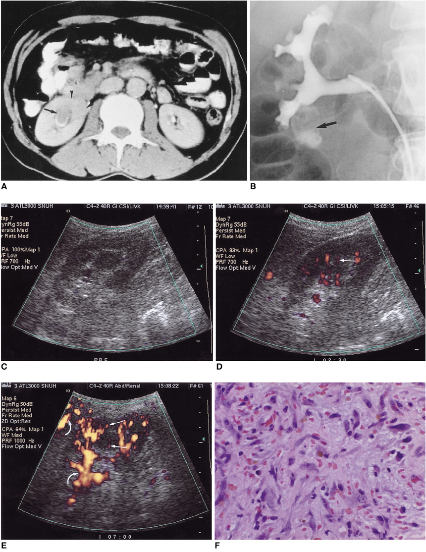
Fig. 1 A 60-year-old man with gross hematuria first noted ten days earlier. A. Delayed enhanced CT image shows a well-circumscribed, right renal mass (arrow) involving the lower pole calyx. Areas of low attenuation (arrowheads) are present in adjacent renal parenchyma. B. Retrograde pyelography revealed a dilated lower pole calyx with irregular filling defect (arrow). C. Gray-scale US demonstrated a round, homogeneously hypoechoic lesion, 1.6 cm in diameter, at the lower pole of the right kidney (not shown). Unenhanced power Doppler US revealed no definite tumoral vascularity. D. Harmonic power Doppler US image obtained 150 seconds after the injection of contrast agent shows dot-like, linear power Doppler US signals within the mass (arrow). These were not depicted by unenhanced power Doppler US (C). E. Conventional power Doppler US image obtained 7 minutes after injection also shows dot-like, linear power Doppler US signals. Compared with harmonic power Doppler US, severe blooming artifacts around and within the kidney are noted (curved arrows). F. Photomicrograph of a histologic specimen shows marked proliferation of myofibroblasts and capillaries with infiltration of lymphocytes, plasma cells and macrophages. Mitotic activity was low, and cellular atypism was minimal.
Reference
-
1. Horiuchi R, Uchida T, Kojima T, et al. Inflammatory pseudotumor of the liver: Clinicopathologic study and review of the literature. Cancer. 1990. 65:1583–1590.2. Bell ND, Gavras JN, Donnell CA, Rodning CB. Renal inflammatory pseudotumor. South Med J. 1998. 91:1050–1053.3. Vujanic GM, Berry PJ, Frank JD. Inflammatory pseudotumor of the kidney with extensive metaplastic bone. Pediatr Pathol. 1992. 12:557–561.4. Young RH, Scully RE. Pseudosarcomatous lesions of the urinary bladder, prostate gland, and urethra: A report of three cases and review of the literature. Arch Pathol Lab Med. 1987. 111:354–358.5. Jones EC, Clement PB, Young RH. Inflammatory pseudotumor of the urinary bladder: A clinicopathological, immunohistochemical, ultrastructural, and flow cytometric study of 13 cases. Am J Surg Pathol. 1993. 17:264–274.6. Materne R, Van Beers BE, Gigot JF, et al. Inflammatory pseudotumor of the liver: MRI with mangafodipir trisodium. J Comput Assist Tomogr. 1998. 22:82–84.7. Abehsera M, Vilgrain V, Belghiti J, Flejou JF, Nahum H. Inflammatory pseudotumor of the liver: radiologic-pathologic correlation. J Comput Assist Tomogr. 1995. 19:80–83.8. Nam KJ, Kang HK, Lim JH. Inflammatory pseudotumor of the liver: CT and sonographic findings. AJR. 1996. 167:485–487.9. Kim AY, Kim SH, Kim YJ, et al. Contrast-enhanced power Doppler sonography for the differentiation of cystic renal lesions: preliminary study. J Ultrasound Med. 1999. 18:581–588.10. Choi BI, Kim TK, Han JK, et al. Vascularity of hepatocellular carcinoma: assessment with contrast-enhanced second-harmonic versus conventional power Doppler US. Radiology. 2000. 214:381–386.
