Successful Management with Glue Injection of Arterial Rupture Seen during Embolization of an Arteriovenous Malformation Using a Flow-Directed Catheter: A Case Report
- Affiliations
-
- 1Department of Diagnostic Radiology, Wallace Memorial Baptist Hospital, Pusan, Korea. cinemani@nownuri.net
- KMID: 1378946
- DOI: http://doi.org/10.3348/kjr.2000.1.4.208
Abstract
- We present a case in which an arterial rupture occurring during embolization of an arteriovenous malformation of the left occipital lobe with a flow-directed micro-catheter,was successfully sealed with a small amount of glue. We navigated a 1.8-Fr Magic catheter through the posterior cerebral artery, and during superse-lective test injection, extravasation was observed at the parieto-occipital branch. The catheter was not removed and the perforation site was successfully sealed with a small amount of glue injected through the same catheter. Prompt recogni-tion and closure of the perforation site is essential for good prognosis.
Keyword
MeSH Terms
Figure
-
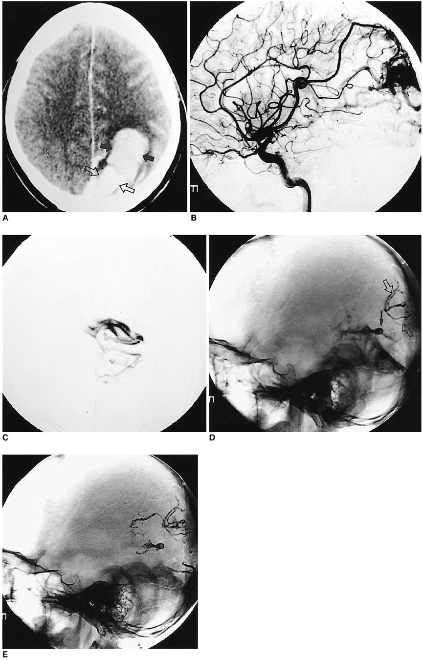
Fig. 1 A. Axial enhanced CT reveals a left occipital arteriovenous malformation (open arrows). A hematoma (arrows) is seen in the left parietal lobe. No feeding artery appears in this section. B. Lateral angiograms of the left internal carotid artery disclose an occipital arteriovenous malformation fed by the left posterior parietal artery and branches of the posterior cerebral artery. C. Anteroposterior test injection shows contrast extravasation into the subarachnoid space at the point at which the parieto-occipital branch changes direction, the site of the microcatheter tip, approximately 1.5 cm from the nidus. D. Plain lateral view shows a mixture of histoacryl and lipiodol after occlusion of the arterial rupture with glue (arrow), and change in the position and shape of the initial extravasation of contrast medium is seen. Note the presence of glue cast arising from initial embolization of the left posterior parietal artery (open arrows). E. Plain lateral view obtained 139 minutes after initial contrast extravasation shows no evidence of further extravasation.
Reference
-
1. Wikholm G. Occlusion of cerebral arteriovenous malformations with n-butyl cyanoacrylate is permanent. AJNR. 1995. 16:479–482.2. Halbach V, Higashida R, Dowd C, Barnwell S, Hieshima G. Management of vascular perforations that occur during neurointerventional procedures. AJNR. 1991. 12:319–327.3. Purdy PD, Samson D, Batjer HH, et al. Preoperative embolization of central arteriovenous malformation with polyvinyl alcohol particles: experience in 51 adults. AJNR. 1990. 11:501–510.4. Dion JE, Duckwiler GR, Lylyk P, Vinuela F, Bentson J. Progressive suppleness pursil catheter: a new tool for superselective angiography and embolization. AJNR. 1989. 10:1068–1070.5. Aletich VA, Debrun GM, Koenigsberg R, Ausman JI, Charbel F, Dujovny M. Arteriovenous malformation nidus catheterization with hydrophilic wire and flow-directed catheter. AJNR. 1997. 18:929–935.6. Lasjaunias P, Berenstein A. Surgical Neuroangiography. 1987. 2nd ed. New York: Springer-Verlag;96–97.
- Full Text Links
-
- Actions
-
Cited
- CITED
-
- Close
- Share
-
- Similar articles
-
- Embolization of Uterine Arteriovenous Malformation
- A Case of Arteriovenous Malformation of the Nasal Tip
- Arteriovenous Fistula at Scalp: Rapid Progression After Embolization of Contralateral Facial Arteriovenous Malformation
- Dural Arteriovenous Fistula of Jugular Foramen with Subarachnoid Hemorrhage : Selective Transarterial Embolization
- A Case of Congenital Renal Arteriovenous Malformation with Severe Gross Hematuria
