EUS Elastography: Advances in Diagnostic EUS of the Pancreas
- Affiliations
-
- 1Department of Internal Medicine, Institute for Digestive Research, Soonchunhyang University College of Medicine, Seoul 140-887, Korea. ydcho@schmc.ac.kr
- KMID: 1372862
- DOI: http://doi.org/10.3348/kjr.2012.13.S1.S12
Abstract
- Elastography is an imaging modality for the evaluation of tissue stiffness, which has been used for the analysis of superficial organs, such as those of the breast and prostate. The measurement of tissue elasticity has been reported to be useful for the diagnosis and differentiation of tumors, which are stiffer than normal tissues. Endoscopic ultrasonography elastography (EUS-EG) is a promising imaging technique with a high degree of accuracy for the differential diagnosis of solid pancreatic tumors. Recent introduction of second generation EUS-EG allows for the quantitative analysis of tissue stiffness. Here, we review our knowledge and preliminary experience with the use of EUS-elastography for the diagnosis of pancreatic disease.
MeSH Terms
Figure
-

Fig. 1 Principles of real-time tissue elastography. When model in which hard springs and soft springs are one-dimensionally connected is compressed, hard springs are negligibly deformed, but soft springs are markedly deformed. Same type of information is provided when sound wave passes through tissues that vary in hardness. This difference in deformation results in differences in displacement among areas, and amount of distortion obtained by spatial differentiation of this displacement distribution provides elasticity information.
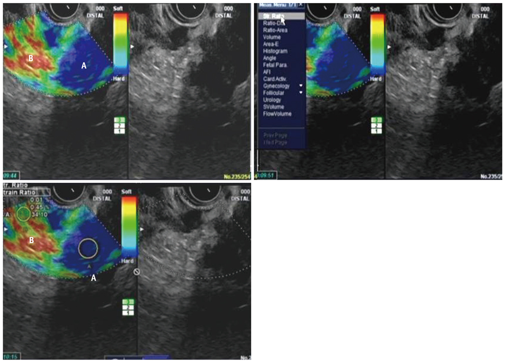
Fig. 2 Quantitative analysis of pancreatic cancer using EUS elastography. Two different areas (A and B) from region of interest were selected for quantitative elastographic analysis. Area A is representative area of mass and included the biggest possible area of the tumor. Area B refers to soft (red) peripancreatic reference area outside tumor. Quotient B/A (strain ratio) is considered as measure of elastographic evaluation.

Fig. 3 EUS elastography of normal pancreas EUS. Elastographic imaging of normal pancreas is characterized by uniform, homogenous green color distribution (representing intermediate stiffness).
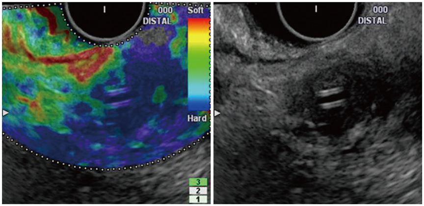
Fig. 4 EUS elastography of pancreatic cancer. Elastographic imaging of pancreatic cancer is characterized by heterogeneous blue color distribution (representing hard stiffness).
Cited by 2 articles
-
Endoscopic Ultrasound Elastography for the Pancreas in Korea: A Preliminary Single Center Study
Tae Hee Lee, Young Deok Cho, Sang-Woo Cha, Joo Young Cho, Jae Young Jang, Soung Won Jeong, Hyun Jong Choi, Jong Ho Moon
Clin Endosc. 2013;46(2):172-177. doi: 10.5946/ce.2013.46.2.172.Primary Fallopian Tube Carcinoma Diagnosed with Endoscopic Ultrasound Elastography with Fine Needle Biopsy
Eui Bae Kim, Tae Hee Lee, Jeong Sig Kim, In Ho Choi
Clin Endosc. 2014;47(5):464-468. doi: 10.5946/ce.2014.47.5.464.
Reference
-
1. Itoh A, Ueno E, Tohno E, Kamma H, Takahashi H, Shiina T, et al. Breast disease: clinical application of US elastography for diagnosis. Radiology. 2006. 239:341–350.2. Cochlin DL, Ganatra RH, Griffiths DF. Elastography in the detection of prostatic cancer. Clin Radiol. 2002. 57:1014–1020.3. Krouskop TA, Wheeler TM, Kallel F, Garra BS, Hall T. Elastic moduli of breast and prostate tissues under compression. Ultrason Imaging. 1998. 20:260–274.4. Giovannini M, Hookey LC, Bories E, Pesenti C, Monges G, Delpero JR. Endoscopic ultrasound elastography: the first step towards virtual biopsy? Preliminary results in 49 patients. Endoscopy. 2006. 38:344–334.5. Saftoiu A, Vilman P. Endoscopic ultrasound elastography--a new imaging technique for the visualization of tissue elasticity distribution. J Gastrointestin Liver Dis. 2006. 15:161–165.6. Janssen J, Schlörer E, Greiner L. EUS elastography of the pancreas: feasibility and pattern description of the normal pancreas, chronic pancreatitis, and focal pancreatic lesions. Gastrointest Endosc. 2007. 65:971–978.7. Giovannini M, Thomas B, Erwan B, Christian P, Fabrice C, Benjamin E, et al. Endoscopic ultrasound elastography for evaluation of lymph nodes and pancreatic masses: a multicenter study. World J Gastroenterol. 2009. 15:1587–1593.8. Iglesias-Garcia J, Larino-Noia J, Abdulkader I, Forteza J, Dominguez-Munoz JE. EUS elastography for the characterization of solid pancreatic masses. Gastrointest Endosc. 2009. 70:1101–1108.9. Hirche TO, Ignee A, Barreiros AP, Schreiber-Dietrich D, Jungblut S, Ott M, et al. Indications and limitations of endoscopic ultrasound elastography for evaluation of focal pancreatic lesions. Endoscopy. 2008. 40:910–917.10. Ophir J, Céspedes I, Ponnekanti H, Yazdi Y, Li X. Elastography: a quantitative method for imaging the elasticity of biological tissues. Ultrason Imaging. 1991. 13:111–134.11. Iglesias-Garcia J, Larino-Noia J, Abdulkader I, Forteza J, Dominguez-Munoz JE. Quantitative endoscopic ultrasound elastography: an accurate method for the differentiation of solid pancreatic masses. Gastroenterology. 2010. 139:1172–1180.12. Byrne MF, Jowell PS. Gastrointestinal imaging: endoscopic ultrasound. Gastroenterology. 2002. 122:1631–1648.13. Kaufman AR, Sivak MV Jr. Endoscopic ultrasonography in the differential diagnosis of pancreatic disease. Gastrointest Endosc. 1989. 35:214–219.14. Giovannini M. Contrast-enhanced endoscopic ultrasound and elastosonoendoscopy. Best Pract Res Clin Gastroenterol. 2009. 23:767–779.
- Full Text Links
-
- Actions
-
Cited
- CITED
-
- Close
- Share
-
- Similar articles
-
- Role of contrast-enhanced harmonic endoscopic ultrasonography (EUS) and EUS elastography in pancreatic lesions
- Interventional Endoscopic Ultrasonography: Present and Future
- Endoscopic Ultrasound in Gastroenteropancreatic Neuroendocrine Tumors
- Endoscopic Ultrasound Images of Normal Anatomy of the Pancreas and Biliary Tree
- Diagnostic Endoscopic Ultrasound: Technique, Current Status and Future Directions
