Value of CT in the Discrimination of Fatal from Non-Fatal Stercoral Colitis
- Affiliations
-
- 1Division of Emergency and Critical Care Radiology, Department of Medical Imaging and Intervention, Chang Gung Memorial Hospital, Chang Gung University, Taoyuan 333, Taiwan. ycwong@adm.cgmh.org.tw
- 2Division of Trauma and Emergency, Department of Surgery, Chang Gung Memorial Hospital, Chang Gung University, Taoyuan 333, Taiwan.
- 3Department of Pathology, Chang Gung Memorial Hospital, Chang Gung University, Taoyuan 333, Taiwan.
- KMID: 1372847
- DOI: http://doi.org/10.3348/kjr.2012.13.3.283
Abstract
OBJECTIVE
Clinical presentation and physical signs may be unreliable in the diagnosis of stercoral colitis (SC). This study evaluates the value of computed tomography (CT) in distinguishing fatal from non-fatal SC.
MATERIALS AND METHODS
Ten patients diagnosed as SC were obtained from inter-specialist conferences. Additional 13 patients with suspected SC were identified via the Radiology Information System (RIS). These patients were divided into two groups; fatal and non-fatal SCs. Their CT images are reviewed by two board-certified radiologists blinded to the clinical data and radiographic reports.
RESULTS
SC occurred in older patients and displayed no gender predisposition. There was significant correlation between fatal SC and CT findings of dense mucosa (p = 0.017), perfusion defects (p = 0.026), ascites (p = 0.023), or abnormal gas (p = 0.033). The sensitivity, specificity, and accuracy of dense mucosa were 71%, 86%, and 81%, respectively. These figures were 75%, 79%, and 77% for perfusion defects; 75%, 80%, and 78% for ascites; and 50%, 93%, and 78% for abnormal gas, respectively. Each CT sign of mucosal sloughing and pericolonic abscess displayed high specificity of 100% and 93% for diagnosing fatal SC, respectively. However, this did not reach statistical significance in diagnosing fatal SC.
CONCLUSION
CT appears to be valuable in discriminating fatal from non-fatal SC.
Keyword
MeSH Terms
-
Adult
Aged
Aged, 80 and over
Chi-Square Distribution
Colitis/mortality/*radiography
Contrast Media/diagnostic use
Diagnosis, Differential
Fecal Impaction/mortality/*radiography
Female
Humans
Male
Middle Aged
Retrospective Studies
Risk Factors
Sensitivity and Specificity
Statistics, Nonparametric
Tomography, X-Ray Computed/*methods
Figure
-
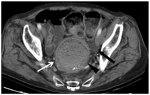
Fig. 1 Enhanced abdomen CT for 71-year-old woman with non-fatal SC demonstrating wall thickening (arrowhead) at recto-sigmoid colon, discontinuation of enhanced mucosa that indicates perfusion defect (white arrow). Regional ascites accumulation (black arrow) is also seen. SC = stercoral colitis
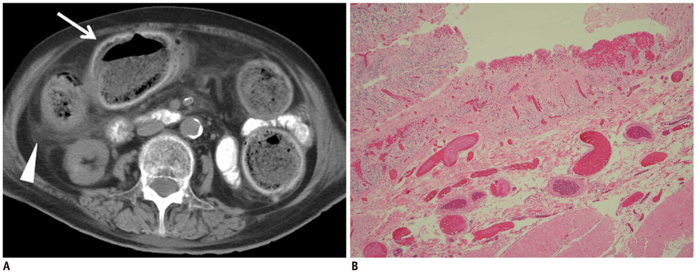
Fig. 2 Fatal stercoral colitis in 79-year old woman. A. Un-enhanced CT showing dense rim conforming to wall of sigmoid colon impressed as dense mucosa (white arrow). Increased streaky pericolic infiltration (arrowhead) is also seen. B. Note presence of mucosal hemorrhage and marked submucosal congestion of tissue specimen.
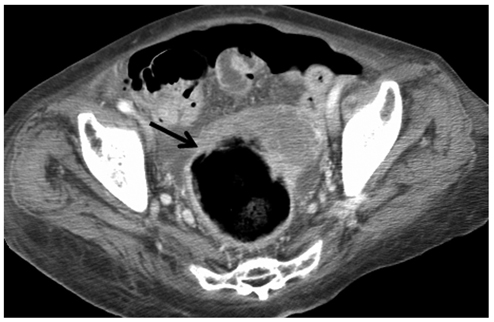
Fig. 3 Enhanced abdomen CT for 88-year-old woman with fatal SC revealing mucosal slip (arrow) sloughing out from wall of distended colon. Surgical resection confirmed SC with gangrene mucosa in sloughing. SC = stercoral colitis
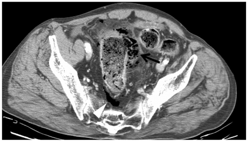
Fig. 4 Enhanced CT for 87-year-old man with non-fatal SC revealing confined mottled substance abutting sigmoid colon (arrow), indicative of perisigmoid abscess formation. Extra-luminal gas (white arrowhead) around the sigmoid colon is also seen. SC = stercoral colitis
Reference
-
1. Yano T, Wakabayashi H, Yachida S, Okano K, Izuishi K, Suzuki Y. A stercoral perforation of the colon with an obvious faecal mass diagnosed by computed tomography. ANZ J Surg. 2008. 78:214–215.2. Wald A. Management and prevention of fecal impaction. Curr Gastroenterol Rep. 2008. 10:499–501.3. Sharma M, Agrawal A. Case report: Stercoral sigmoid colonic perforation with fecal peritonitis. Indian J Radiol Imaging. 2010. 20:126–128.4. Zissin R, Hertz M, Osadchy A, Even-Sapir E, Gayer G. Abdominal CT findings in nontraumatic colorectal perforation. Eur J Radiol. 2008. 65:125–132.5. Serpell JW, Nicholls RJ. Stercoral perforation of the colon. Br J Surg. 1990. 77:1325–1329.6. Haddad R, Bursle G, Piper B. Stercoral perforation of the sigmoid colon. ANZ J Surg. 2005. 75:244–246.7. Wu CH, Wang LJ, Wong YC, Huang CC, Chen CC, Wang CJ, et al. Necrotic stercoral colitis: importance of computed tomography findings. World J Gastroenterol. 2011. 17:379–384.8. Ouaïssi M, Sielezneff I, Benoist S, Pirró N, Cretel E, Chaix JB, et al. Lethal fecaloma. J Am Geriatr Soc. 2007. 55:965–967.9. Rozenblit AM, Cohen-Schwartz D, Wolf EL, Foxx MJ, Brenner S. Case reports. Stercoral perforation of the sigmoid colon: computed tomography findings. Clin Radiol. 2000. 55:727–729.10. Maurer CA, Renzulli P, Mazzucchelli L, Egger B, Seiler CA, Büchler MW. Use of accurate diagnostic criteria may increase incidence of stercoral perforation of the colon. Dis Colon Rectum. 2000. 43:991–998.11. Heffernan C, Pachter HL, Megibow AJ, Macari M. Stercoral colitis leading to fatal peritonitis: CT findings. AJR Am J Roentgenol. 2005. 184:1189–1193.12. Kim MJ, Park SH, Lee SS, Byeon JS, Choi EK, Kim JH, et al. Efficacy of barium-based fecal tagging for CT colonography: a comparison between the use of high and low density barium suspensions in a Korean population - a preliminary study. Korean J Radiol. 2009. 10:25–33.13. Frager DH, Baer JW. Role of CT in evaluating patients with small-bowel obstruction. Semin Ultrasound CT MR. 1995. 16:127–140.14. Furukawa A, Kanasaki S, Kono N, Wakamiya M, Tanaka T, Takahashi M, et al. CT diagnosis of acute mesenteric ischemia from various causes. AJR Am J Roentgenol. 2009. 192:408–416.15. Boudiaf M, Soyer P, Terem C, Pelage JP, Maissiat E, Rymer R. Ct evaluation of small bowel obstruction. Radiographics. 2001. 21:613–624.16. Rha SE, Ha HK, Lee SH, Kim JH, Kim JK, Kim JH, et al. CT and MR imaging findings of bowel ischemia from various primary causes. Radiographics. 2000. 20:29–42.17. Lohrmann C, Ghanem N, Pache G, Makowiec F, Kotter E, Langer M. CT in acute perforated sigmoid diverticulitis. Eur J Radiol. 2005. 56:78–83.18. Miki T, Ogata S, Uto M, Nakazono T, Urata M, Ishibe R, et al. Multidetector-row CT findings of colonic perforation: direct visualization of ruptured colonic wall. Abdom Imaging. 2004. 29:658–662.19. Kim SW, Shin HC, Kim IY, Kim YT, Kim CJ. CT findings of colonic complications associated with colon cancer. Korean J Radiol. 2010. 11:211–221.20. Rubesin SE, Levine MS. Radiologic diagnosis of gastrointestinal perforation. Radiol Clin North Am. 2003. 41:1095–1115. v21. Yeung KW, Chang MS, Hsiao CP, Huang JF. CT evaluation of gastrointestinal tract perforation. Clin Imaging. 2004. 28:329–333.22. Hainaux B, Agneessens E, Bertinotti R, De Maertelaer V, Rubesova E, Capelluto E, et al. Accuracy of MDCT in predicting site of gastrointestinal tract perforation. AJR Am J Roentgenol. 2006. 187:1179–1183.23. Maniatis V, Chryssikopoulos H, Roussakis A, Kalamara C, Kavadias S, Papadopoulos A, et al. Perforation of the alimentary tract: evaluation with computed tomography. Abdom Imaging. 2000. 25:373–379.24. Yoo HJ, Kim SH, Lee JM, Kim MA, Han JK, Choi BI. The association of anisakiasis in the ascending colon with sigmoid colon cancer: CT colonography findings. Korean J Radiol. 2008. 9:Suppl. S56–S60.25. Sood BP, Rezai S, Parker D. Unusual radiological appearance of a faecaloma. Australas Radiol. 2007. 51 Spec No:B161–B164.26. Huang WS, Wang CS, Hsieh CC, Lin PY, Chin CC, Wang JY. Management of patients with stercoral perforation of the sigmoid colon: report of five cases. World J Gastroenterol. 2006. 12:500–503.27. Patel VG, Kalakuntla V, Fortson JK, Weaver WL, Joel MD, Hammami A. Stercoral perforation of the sigmoid colon: report of a rare case and its possible association with nonsteroidal anti-inflammatory drugs. Am Surg. 2002. 68:62–64.28. Chen JH, Shen WC. Rectal carcinoma with stercoral ulcer perforation. Hepatogastroenterology. 2000. 47:1018–1019.
