Thumb Duplication: Concepts and Techniques
- Affiliations
-
- 1Department of Hand Surgery and Peripheral Nerve Surgery, University of Sydney, Royal North Shore Hospital, St. Leonards and Department of Hand Surgery, University of Sydney, Children's Hospital at Westmead, Westmead, Australia. mtonkin@surgery.usyd.edu.au
- KMID: 1245395
- DOI: http://doi.org/10.4055/cios.2012.4.1.1
Abstract
- Within the Oberg, Manske, Tonkin (OMT) classification, thumb duplications are a failure of formation and/or differentiation affecting the radial-ulnar axis of the hand plate. The Wassel description of seven types of thumb duplication provides a good structure from which an approach to management is based. The aim of surgical reconstruction is to obtain a stable, mobile thumb of adequate size and appropriate shape. The most common form of reconstruction is removal of the lesser digit and reconstruction of the dominant digit. Surgical techniques address the problems of deviation, instability and lack of size. The disadvantages of the Bilhaut-Cloquet procedure, these being joint stiffness and a nail ridge, may be lesser concerns when reconstruction of one digit will not create a satisfactory thumb of adequate mobility, stability, alignment and size. Complicated problems of triphalangism, triplication, ulnar dimelia and the rare circumstance in which neither of the duplicated thumbs may be adequately reconstructed present specific challenges which demand alternative techniques.
Keyword
MeSH Terms
Figure
-
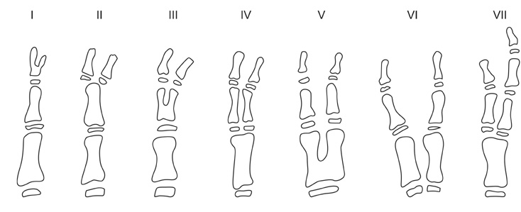
Fig. 1 The Wassel classification of thumb duplication. Reprinted with permission from Springer-Verlag.3)
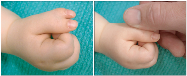
Fig. 2 Appearance of the dominant thumb when the examiner's hand conceals the radial duplicate.
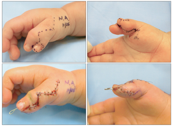
Fig. 3 Elevation of a neurovascular island flap from the radial aspect of the radial thumb to improve the contour of the reconstructed thumb.
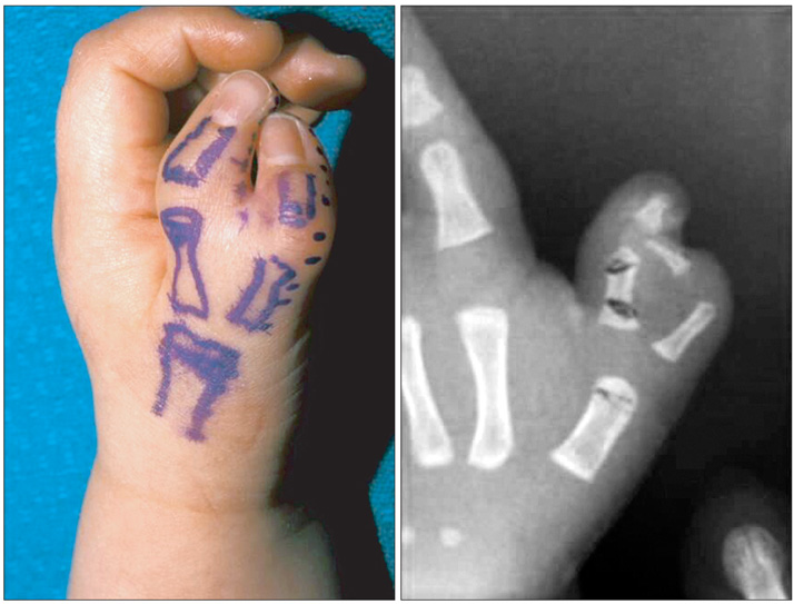
Fig. 4 Wassel type IV thumb duplication with divergence of proximal phalanges and convergence of distal phalanges.
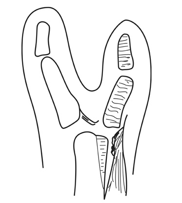
Fig. 5 Diagram of capsular-periosteal flap for metacarpophalangeal joint radial collateral ligament reconstruction; and longitudinal osteotomy to remove redundant articular surface of the metacarpal head.

Fig. 6 Distal bifurcation of the flexor pollicis longus with eccentric insertions into the terminal phalanges.

Fig. 7 Connections between the flexor pollicis longus (white arrow) and the extrinsic extensors (black arrow).

Fig. 8 Corrective osteotomies at the neck of the proximal phalanx (white arrow) and the neck of the metacarpal (black arrow).
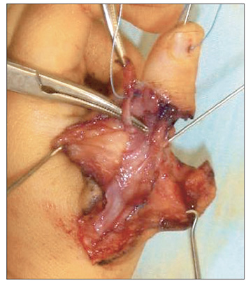
Fig. 9 Tendon realignment and pulley reconstruction.

Fig. 10 Reconstruction of the metacarpophalangeal joint radial collateral ligament with a capsular-periosteal flap. Soft tissue from the base of the proximal phalanx is sutured to the metacarpal neck (white arrow). Radial collateral ligament and thenar musculature is oversewn into the ligament reconstruction (black arrow).

Fig. 11 The "elephant trunk" sign of global metacarpophalangeal joint instability.
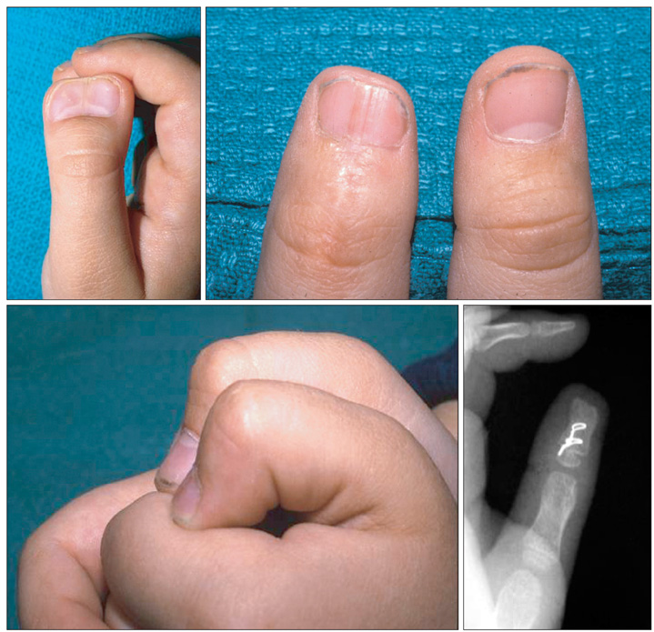
Fig. 12 Preoperative and postoperative views of a Wassel type I with equal size and shape of duplicate parts.

Fig. 13 Diagram of excision of triangular fragments of bone and cartilage from the proximal phalangeal head to decompress the terminal phalangeal reconstruction.
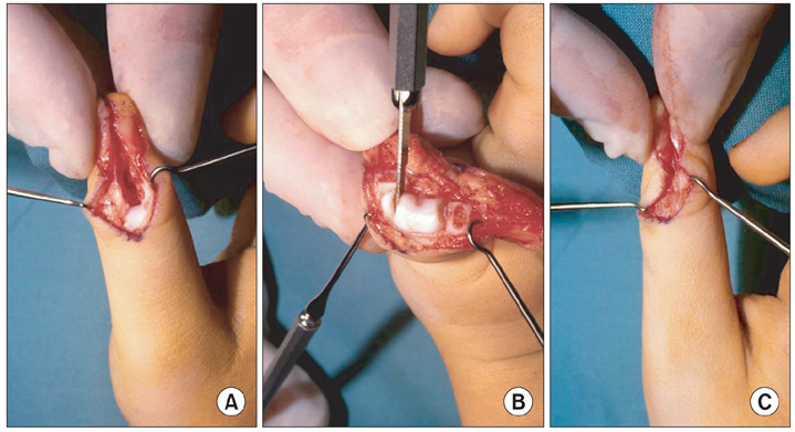
Fig. 14 (A) Inability to appose the two parts of the terminal phalanx along their whole length. (B) Removal of triangular wedge of cartilage and bone from the proximal phalangeal head bilaterally. (C) Easy apposition of terminal phalangeal components.
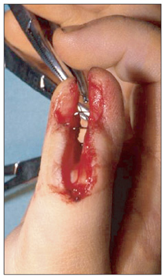
Fig. 15 Two fine interosseous wires are placed at defined distances distal to the physes.

Fig. 16 Significant instability and deviation of a type III thumb duplication.

Fig. 17 Bilhaut-Cloquet reconstruction of a type IV thumb duplication with good carpometacarpal and metacarpophalangeal joint motion, restricted interphalangeal joint motion, good size and appearance and minimal ridging.
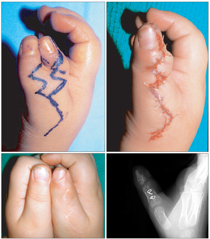
Fig. 18 Modified Bilhaut-Cloquet procedure.

Fig. 19 Preoperative appearance prior to combination of acral transposition of the distal part of the ulnar thumb to the radial thumb with shortening and fusions.
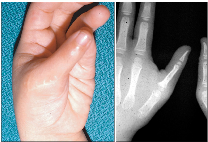
Fig. 20 Postoperative result from the case shown in Fig. 19.

Fig. 21 Preoperative appearance and X-ray of a thumb duplication.

Fig. 22 Postoperative appearance and X-ray following pollicisation of the dominant digit in the case shown in Fig. 21.

Fig. 23 Shortening, rotational metacarpal osteotomy and interphalangeal fusion with first web reconstruction of the optimal digit in a case of bilateral thumb triplication.
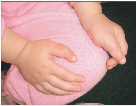
Fig. 24 Postoperative appearance from the case shown in Fig. 23.
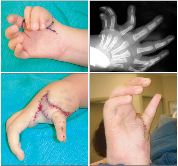
Fig. 25 Pre- and postoperative appearance in a case of ulnar dimelia in which optimal appearance and function were obtained by reducing the number of digits to four.
Reference
-
1. Swanson AB. A classification for congenital limb malformations. J Hand Surg Am. 1976. 1(1):8–22.
Article2. Oberg KC, Feenstra JM, Manske PR, Tonkin MA. Developmental biology and classification of congenital anomalies of the hand and upper extremity. J Hand Surg Am. 2010. 35(12):2066–2076.
Article3. Wassel HD. The results of surgery for polydactyly of the thumb. Clin Orthop Relat Res. 1969. 64:175–193.4. Kozin SH. Wolfe SW, Hotchkiss RN, Pederson WC, Kozin SH, editors. Deformities of the thumb. Green's operative hand surger. 2011. 6th ed. Philadelphia, PA: Elsevier;1383–1394.
Article5. Bilhaut M. Guerison d'un pouce bifide par un nouveau procede operatoire. Congr Fr Chir. 1890. 4:576–580.6. Miura T. Duplicated thumb. Plast Reconstr Surg. 1982. 69(3):470–481.
Article7. Horii E, Nakamura R, Sakuma M, Miura T. Duplicated thumb bifurcation at the metacarpophalangeal joint level: factors affecting surgical outcome. J Hand Surg Am. 1997. 22(4):671–679.
Article8. Gupta A, Kay SP, Scheker L. The growing hand. 2000. London, UK: Mosby;244–252.9. Ogino T, Ishii S, Minami M. Radially deviated type of thumb polydactyly. J Hand Surg Br. 1988. 13(3):315–319.
Article10. Dobyns JH, Lipscomb PR, Cooney WP. Management of thumb duplication. Clin Orthop Relat Res. 1985. (195):26–44.
Article11. Tada K, Yonenobu K, Tsuyuguchi Y, Kawai H, Egawa T. Duplication of the thumb: a retrospective review of two hundred and thirty-seven cases. J Bone Joint Surg Am. 1983. 65(5):584–598.
Article12. Naasan A, Page RE. Duplication of the thumb: a 20-year retrospective review. J Hand Surg Br. 1994. 19(3):355–360.13. Manske PR. Treatment of duplicated thumb using a ligamentous/periosteal flap. J Hand Surg Am. 1989. 14(4):728–733.
Article14. Tonkin MA, Rumball KM. The Bilhaut-Cloquet procedure revisited. Hand Surg. 1997. 2(1):67–74.
Article15. Tonkin MA, Bulstrode NW. The Bilhaut-Cloquet procedure for Wassel types III, IV and VII thumb duplication. J Hand Surg Eur Vol. 2007. 32(6):684–693.
Article
- Full Text Links
-
- Actions
-
Cited
- CITED
-
- Close
- Share
-
- Similar articles
-
- Evolutionary and Comparative Genomics to Drive Rational Drug Design, with Particular Focus on Neuropeptide Seven-Transmembrane Receptors
- Heterodigital flap as a solution for a thumb defect: a case report
- A Case of Tubular Duplication of the Esophagus
- Clinical experiences in Thumb reconstruction
- The Effect of Thumb Position on Median-Radial Latency Difference
