Korean J Urol.
2008 Oct;49(10):953-956. 10.4111/kju.2008.49.10.953.
Renal Intracystic Massive Hemorrhage after Blunt Trauma
- Affiliations
-
- 1Department of Urology, Inje University College of Medicine, Busan, Korea. ur0kang@hanmail.net
- 2Department of Radiology, Inje University College of Medicine, Busan, Korea.
- 3Department of Pathology, Inje University College of Medicine, Busan, Korea.
- KMID: 1204327
- DOI: http://doi.org/10.4111/kju.2008.49.10.953
Abstract
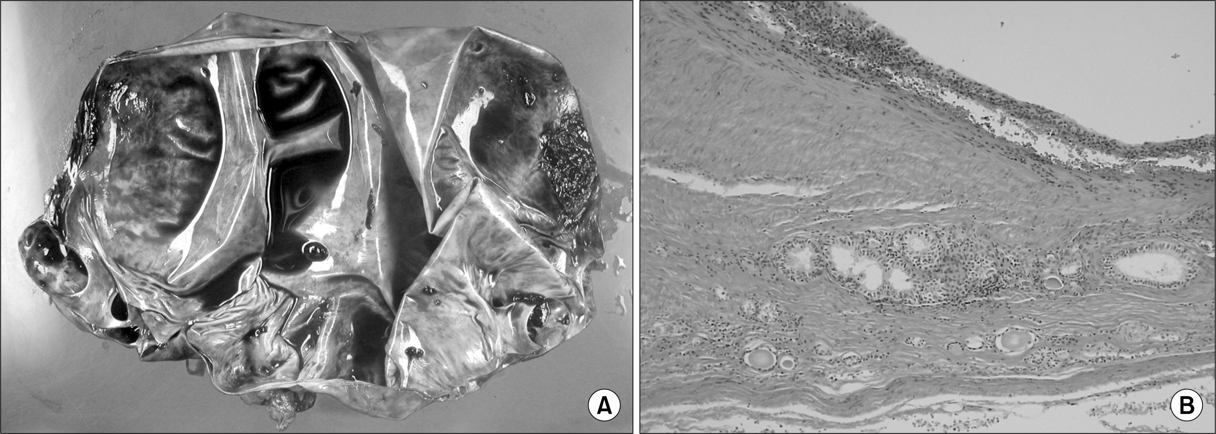
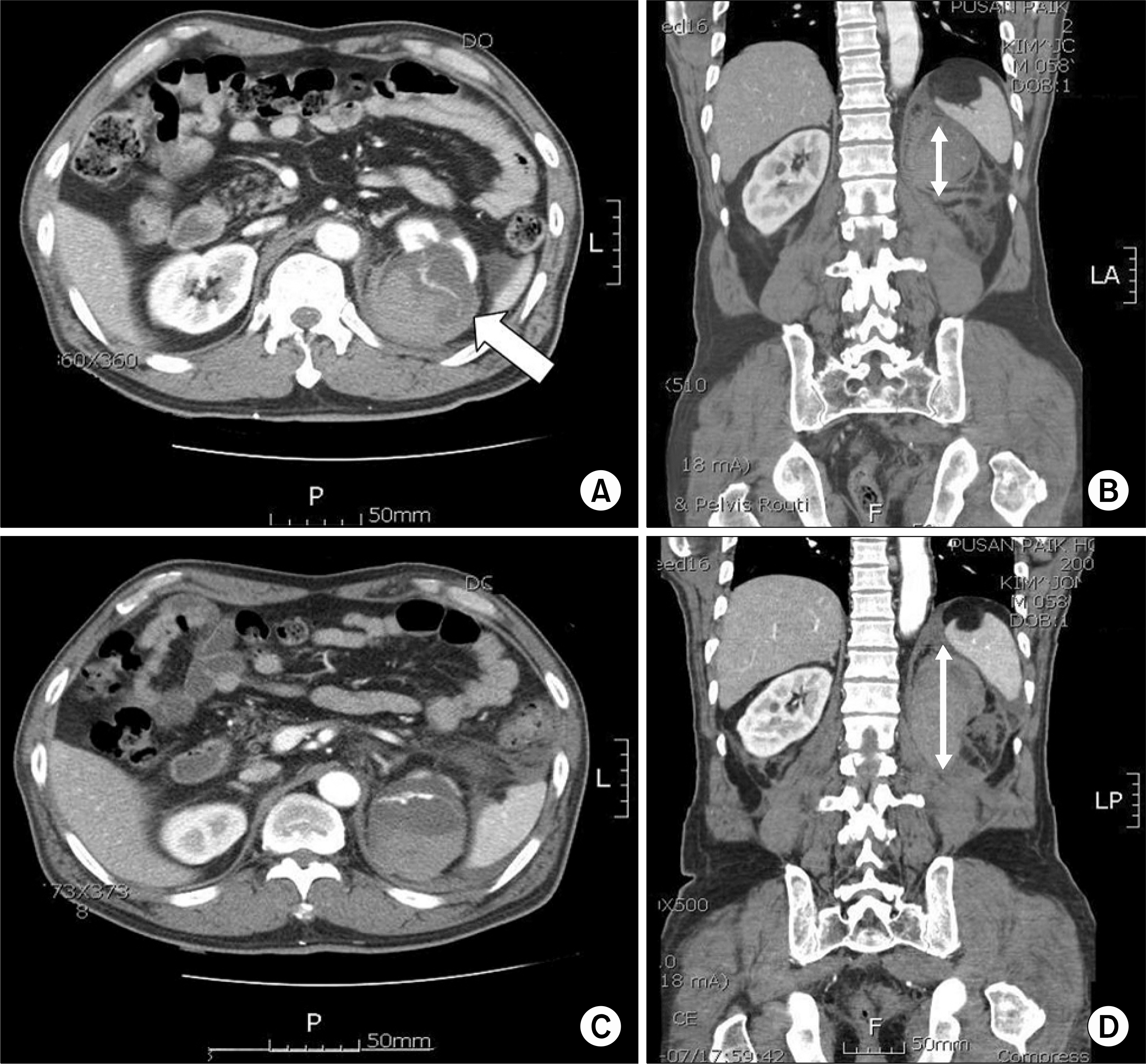
- Spontaneous and post-traumatic renal intracystic hemorrhages are extremely rare, but are a potential danger to patients with cystic kidney disease. We report two cases of post-traumatic intracystic massive hemorrhage in renal cysts. One patient was a 27-year-old male who presented with left flank pain and gross hematuria after slipping on the stairs 2 days previously. The other patient was a 58-year-old male who presented with back pain due to an accident. The circulatory states of the two patients were deteriorated and renal intracystic hemorrhages were detected on computed tomography. One patient underwent a simple nephrectomy and the other patient was treated with arterial embolization. We present two cases of renal intracystic hemorrhage, emphasizing early diagnosis and the treatment of choice.
Keyword
Figure
Reference
-
References
1. McLaughlin AP 3rd, Pfister RC. Spontaneous rupture of renal cysts into the pyelocaliceal system. J Urol. 1975; 113:2–7.
Article2. Hughes CR, Stewart PF Jr, Breckenridge JW. Renal cyst rupture following blunt abdominal trauma: case report. J Trauma. 1995; 38:28–9.3. Rauschmeier H, Jakse G. Traumatic rupture of renal cyst into the retroperitoneum and the pyelocalyceal system. Int Urol Nephrol. 1983; 15:117–9.
Article4. Papanicolaou N, Pfister RC, Yoder IC. Spontaneous and traumatic rupture of renal cysts: diagnosis and outcome. Radiology. 1986; 160:99–103.
Article5. Rainio J, De Giorgio F, Carbone A. Death from renal cyst: spontaneous or traumatic rupture? Am J Forensic Med Pathol. 2006; 27:193–5.6. Redmond PL, Royle G. Rupture of a calcified renal cyst following blunt abdominal trauma. Br J Radiol. 1985; 58:683–4.
Article7. Lopez Cubillana P, Hita Rosino E, Asensio Egea L, Rigabert Montiel M, Hita Villaplana G, Perez Albacete M. Wunderich syndrome. Review of its diagnosis and therapy. Report of seven cases. Actas Urol Esp. 1995; 19:772–6.8. Hwang HH, Cheon SH, Moon KH, Lee SK, Choo HS, Hwang JC, et al. Renal ruptures with active bleeding treated with emergency selective renal arterial embolizaton. Korean J Urol. 2008; 49:177–81.
- Full Text Links
- Actions
-
Cited
- CITED
-
- Close
- Share
- Similar articles
-
- Arachnoid Cyst with Spontaneous Intracystic Hemorrhage and Chronic Subdural Hematoma
- A Case of Control of Renal Hemorrhage by Selective Renal Arterial Embolization
- Intracystic Hemorrhage of an Arachnoid Cyst: a Case with Prediagnostic Imaging of an Intact Cyst
- Renal Artery Pseudoaneurysm after Blunt Renal Trauma
- The Significance of Abdominal Ultrasonography as the Initial Diagnostic Method in Blunt Renal Trauma


