Clin Orthop Surg.
2009 Mar;1(1):11-18. 10.4055/cios.2009.1.1.11.
The Relationship between Spinal Stenosis and Neurological Outcome in Traumatic Cervical Spine Injury: An Analysis using Pavlov's Ratio, Spinal Cord Area, and Spinal Canal Area
- Affiliations
-
- 1Department of Orthopedic Surgery, College of Medicine, Research Institute of Clinical Medicine, Chonbuk National University, Jeonju, Korea.
- 2Department of Orthopedic Surgery, Gwangju Veterans Hospital, Gwangju, Korea. alla1013@naver.com
- KMID: 1127909
- DOI: http://doi.org/10.4055/cios.2009.1.1.11
Abstract
-
BACKGROUND: This study examined the relationship between four radiological parameters (Pavlov's ratio, sagittal diameter, spinal cord area, and spinal canal area) in patients with a traumatic cervical spine injury, as well as the correlation between these parameters and the neurological outcome.
METHODS
A total of 212 cervical spinal levels in 53 patients with a distractive-extension injury were examined. The following four parameters were measured: Pavlov's ratio on the plain lateral radiographs, the sagittal diameter, the spinal cord area, and the spinal canal area on the MRI scans. The Pearson correlation coefficients between the parameters at each level and between the levels of each parameter were evaluated. The correlation between the radiological parameters and the spinal cord injury status classified into four categories, A (complete), B (incomplete), C (radiculopathy), and D (normal) was assessed.
RESULTS
The mean Pavlov's ratio, sagittal diameter, spinal cord area and spinal canal area was 0.84, 12.9 mm, 82.8 mm2 and 236.8 mm2, respectively. An examination of the correlation between the radiological spinal stenosis and clinical spinal cord injury revealed an increase in the values of the four radiological parameters from cohorts A to D. Pavlov's ratio was the only parameter showing statistically significant correlation with the clinical status (p = 0.006).
CONCLUSIONS
There was a correlation between the underlying spinal stenosis and the development of neurological impairment after a traumatic cervical spine injury. In addition, it is believed that Pavlov's ratio can be used to help determine and predict the neurological outcome.
Keyword
MeSH Terms
Figure
-
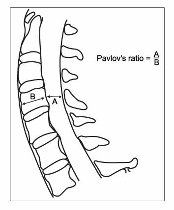
Fig. 1 Pavlov's ratio at each level from C3 to C7.

Fig. 2 The sagittal diameters of the spinal canal were measured at the midvertebra level on the MR T2 sagittal images from C3 to C7.
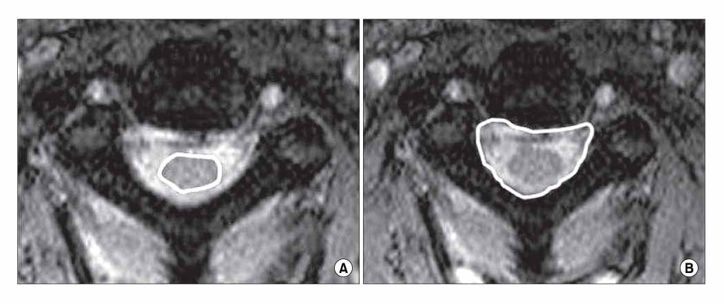
Fig. 3 From T2 axial images, (A) the area of the cord, (B) the area of spinal canal area were measured from C3 to C7.
Reference
-
1. Boden SD, Dodge LD, Bohlman HH, Rechtine GR. Rheumatoid arthritis of the cervical spine. A longterm analysis with predictors of paralysis and recovery. J Bone Joint Surg Am. 1993. 75(9):1282–1297.
Article2. Gore DR. Roentgenographic findings in the cervical spine in asymptomatic persons: a ten-year follow-up. Spine. 2001. 26(22):2463–2466.
Article3. Kang JD, Figgie MP, Bohlman HH. Sagittal measurements of the cervical spine in subaxial fractures and dislocations: An analysis of two hundred and eighty-eight patients with and without neurological deficits. J Bone Joint Surg Am. 1994. 76(11):1617–1628.
Article4. Murone I. The importance of the sagittal diameters of the cervical spinal canal in relation to spondylosis and myelopathy. J Bone Joint Surg Br. 1974. 56(1):30–36.5. Torg JS, Naranja RJ Jr, Pavlov H, Galinat BJ, Warren R, Stine RA. The relationship of developmental narrowing of the cervical spinal canal to reversible and irreversible injury of the cervical spinal cord in football players. J Bone Joint Surg Am. 1996. 78(9):1308–1314.6. Houser OW, Onofrio BM, Miller GM, Folger WN, Smith PL. Cervical spondylotic stenosis and myelopathy: evaluation with computed tomographic myelography. Mayo Clin Proc. 1994. 69(6):557–563.
Article7. Lee CS, Kim YT, Lee CK. Morphological analysis of the cervical spinal cord, dural tube, and spinal canal by Magnetic resonance Imaging in normal korean adults. J Korean Orthop Assoc. 1999. 34(2):265–271.
Article8. Matsuura P, Waters RL, Adkins RH, Rothman S, Gurbani N, Sie I. Comparison of computerized tomography para-meters of the cervical spine in normal control subjects and spinal cord-injured patients. J Bone Joint Surg Am. 1989. 71(2):183–188.
Article9. Pavlov H, Torg JS, Robie B, Jahre C. Cervical spinal stenosis: determination with vertebral body ratio method. Radiology. 1987. 164(3):771–775.
Article10. Penning L, Wilmink JT, van Woerden HH, Knol E. CT myelographic findings in degenerative disorders of the cervical spine: clinical significance. AJR Am J Roentgenol. 1986. 146(4):793–801.
Article11. Okada Y, Ikata T, Yamada H, Sakamoto R, Katoh S. Magnetic resonance imaging study on the results of surgery for cervical compression myelopathy. Spine. 1993. 18(14):2024–2029.
Article12. Lim JK, Wong HK. Variation of the cervical spinal Torg ratio with gender and ethnicity. Spine J. 2004. 4(4):396–401.13. Prasad SS, O'Malley M, Caplan M, Shackleford IM, Pydisetty RK. MRI measurements of the cervical spine and their correlation to Pavlov's ratio. Spine. 2003. 28(12):1263–1268.
Article14. Countee RW, Vijayanathan T. Congenital stenosis of the cervical spine: diagnosis and management. J Natl Med Assoc. 1979. 71(3):257–264.15. Torg JS, Pavlov H, Genuario SE, et al. Neurapraxia of the cervical spinal cord with transient quadriplegia. J Bone Joint Surg Am. 1986. 68(9):1354–1370.
Article16. Blackley HR, Plank LD, Robertson PA. Determining the sagittal dimensions of the canal of the cervical spine. The reliability of ratios of anatomical measurements. J Bone Joint Surg Br. 1999. 81(1):110–112.17. Ono K, Ota H, Tada K, Yamamoto T. Cervical myelopathy secondary to multiple spondylotic protrusion: A clinicopathologic study. Spine. 1977. 2(2):109–125.
Article
- Full Text Links
-
- Actions
-
Cited
- CITED
-
- Close
- Share
-
- Similar articles
-
- Morphological Analysis of the Cervical Spinal Cord, Dural Tube, and Spinal Canal by Magnetic Resonance Imaging in Normal Korean Adults
- The Significance of Space Available for the Spinal cord at the Injured Level in the Lower Cervical Spine Fractures and Dislocations
- The Laminoplasty for Acute Cervical Spinal Cord Injury in Cervical Spondylosis
- Clinical Influence of Cervical Spinal Canal Stenosis on Neurological Outcome after Traumatic Cervical Spinal Cord Injury without Major Fracture or Dislocation
- Soft Tissue Damage in Cervical Spine Extension Injury
