Yonsei Med J.
2007 Oct;48(5):810-817. 10.3349/ymj.2007.48.5.810.
The Relationship between the Left Atrial Volume and the Maximum P-wave and P-wave Dispersion in Patients with Congestive Heart Failure
- Affiliations
-
- 1Department of Cardiology, Inha University College of Medicine, Incheon, Korea. kdhmd@korea.com, kdhmd@inha.ac.kr
- KMID: 1122619
- DOI: http://doi.org/10.3349/ymj.2007.48.5.810
Abstract
- PURPOSE: A maximum P-wave duration (Pmax) of > or = 110msec and a P-wave dispersion (PWD) > or = 40msec are accepted indicators of a disturbance in interatrial conduction and an inhomogeneous propagation of the sinus impulse, respectively. The left atrial (LA) volume has been reported to be strongly associated with a systolic and diastolic dysfunction and is considered to be an index of atrial remodeling. We aimed to investigate the relationship between LA volume and Pmax or PWD in patients with congestive heart failure (CHF). PATIENTS AND METHODS: Sixty-one patients with CHF were enrolled in this study. The study population was classified into four groups: two groups were divided according to the Pmax (> or = 110msec or < 110ms), and the other two groups were formed based on the PWD (> or = 40msec or < 40msec). The left atrial volume index (LAVi) was measured by three-dimensional (3-D) transthoracic echocardiography. The Pmax and PWD were measured from a 12-lead electrocardiogram. RESULTS: There were significant differences in the ejection fraction (EF), diastolic function, and LAVi between patients with a Pmax > or = 110ms or a PWD > or = 40ms and those with a Pmax < 110ms or a PWD < 40ms. The LAVi was independently associated with a disturbance in interatrial conduction and an inhomogeneous propagation of the sinus impulse. The LAVi can be used to identify patients with a disturbance in interatrial conduction and an inhomogeneous propagation of the sinus impulse with reasonably good accuracy. CONCLUSION: We concluded that a disturbance in interatrial conduction and an inhomogenous propagation of the sinus impulse in patients with CHF is associated with an increase in the LA volume and a deleterious systolic and diastolic dysfunction.
MeSH Terms
Figure
-

Fig. 1 On the ROC curve, the AUC of the LAVi for predicting a disturbance in the interatrial conduction was 0.792 (sensitivity 78.0%, specificity 89.5%, positive predictive value 78.1%, negative predictive value 89.5%), and the optimum cut-off point was 48.03 mL/m2 (p < 0.0001). ROC, receiver operating characteristic; AUC, accuracy; LAVi, left atrial volume index.
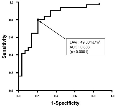
Fig. 2 On the ROC curve, the AUC of LAVi for predicting an inhomogeneous propagation of the sinus impulse was 0.833 (sensitivity 80.6%, specificity 79.3%, positive predictive value 80.6%, negative predictive value 79.3%), and the optimum cut-off point was 49.80 mL/m2 (p < 0.0001). ROC, receiver operating characteristic; AUC, accuracy, LAVi, left atrial volume index.
Reference
-
1. Bayes De Luna A. Electrocardiographic alteration due to atrial pathology. Clinical electrocardiography: a textbook. 1998. New York, NY: Futura Company;169–171.2. Willems JL, Robles de Medina EO, Bernard R, Coumel P, Fisch C, Krikler D, et al. Criteria for intraventricular conduction disturbances and preexcitation. World Health Organizational/International Society and Federation of Cardiology Task Force Ad Hoc. J Am Coll Cardiol. 1985. 5:1261–1275.
Article3. Dilaveris PE, Gialafos EJ, Sideris SK, Theopistou AM, Andrikopoulos GK, Kyriakidis M, et al. Simple electrocardiographic markers for the prediction of paroxysmal idiopathic atrial fibrillation. Am Heart J. 1998. 135:733–738.
Article4. Dilaveris PE, Gialafos EJ, Andrikopoulos GK, Richter DJ, Papanikolaou V, Poralis K, et al. Clinical and electrocardiographic predictors of recurrent atrial fibrillation. Pacing Clin Electrophysiol. 2000. 23:352–358.
Article5. Benjamin EJ, D'Agostino RB, Belanger AJ, Wolf PA, Levy D. Left atrial size and the risk of stroke and death. The Framingham Heart Study. Circulation. 1995. 92:835–841.6. Tsang TS, Barnes ME, Bailey KR, Leibson CL, Montgomery SC, Takemoto Y, et al. Left atrial volume: important risk marker of incident atrial fibrillation in 1665 older men and women. Mayo Clin Proc. 2001. 76:467–475.
Article7. Pritchett AM, Mahoney DW, Jacobsen SJ, Rodeheffer RJ, Karon BL, Redfield MM. Diastolic dysfunction and left atrial volume: a population-based study. J Am Coll Cardiol. 2005. 45:87–92.8. Gottdiener JS, Kitzman DW, Aurigemma GP, Arnold AM, Manolio TA. Left atrial volume, geometry, and function in systolic and diastolic heart failure of persons ≥ 65 years of age (The Cardiovascular Health Study). Am J Cardiol. 2006. 97:83–89.
Article9. Poli S, Barbaro V, Bartolini P, Calcagnini G, Censi F. Prediction of atrial fibrillation from surface ECG: review of methods and algorithms. Ann Ist Super Sanita. 2003. 39:195–203.10. Goyal SB, Spodick DH. Electromechanical dysfunction of the left atrium associated with interatrial block. Am Heart J. 2001. 142:823–827.
Article11. Ramsaran EK, Spodick DH. Electromechanical delay in the left atrium as a consequence of interatrial block. Am J Cardiol. 1996. 77:1132–1134.
Article12. Tsang TS, Gersh BJ, Appleton CP, Tajik AJ, Barnes ME, Bailey KR, et al. Left ventricular diastolic dysfunction as a predictor of the first diagnosed nonvalvular atrial fibrillation in 840 elderly men and women. J Am Coll Cardiol. 2002. 40:1636–1644.
Article13. Tsang TS, Barnes ME, Gersh BJ, Baily KR, Seward JB. Left atrial volume as a morphophysiologic expression of left ventricular diastolic dysfunction and relation to cardiovascular risk burden. Am J Cardiol. 2002. 90:1284–1289.
Article14. Snoeck J, Decoster H, Vrints C, Marchand X, Kahn JC, Verherstraeten M, et al. Predictive value of the P wave at implantation for atrial fibrillation after VVI pacemaker implantation. Pacing Clin Electrophysiol. 1992. 15:2077–2083.
Article15. Shettigar UR, Barry WH, Hultgren HN. P wave analysis in ischaemic heart disease. An echocardiographic, haemodynamic, and angiographic assessment. Br Heart J. 1977. 39:894–899.
Article16. Waggoner AD, Adyanthaya AV, Quinones MA, Alexander JK. Left atrial enlargement. Echocardiographic assessment of electrocardiographic criteria. Circulation. 1976. 54:553–557.
Article17. Sahn DJ, DeMaria A, Kisslo J, Weyman A. Recommendations regarding quantitation in M-mode echocardiography: results of a survey of echocardiographic measurements. Circulation. 1978. 58:1072–1083.
Article18. Schiller NB, Shah PM, Crawford M, DeMaria A, Devereux R, Feigenbaum H, et al. Recommendations for quantitation of the left ventricle by two-dimensional echocardiography. American Society of Echocardiography Committee on Standards, Subcommittee on Quantitation of Two-Dimensional Echocardiograms. J Am Soc Echocardiogr. 1989. 2:358–367.
Article19. Devereux RB, Alonso DR, Lutas EM, Gottlieb GJ, Campo E, Sachs I, et al. Echocardiographic assessment of left ventricular hypertrophy: comparison to necropsy findings. Am J Cardiol. 1986. 57:450–458.
Article20. Senen K, Turhan H, Riza Erbay A, Basar N, Saatci Yasar A, Sahin O, et al. P-wave duration and P-wave dispersion in patients with dilated cardiomyopathy. Eur J Heart Fail. 2004. 6:567–569.
Article21. Greenberg B, Chatterjee K, Parmley WW, Werner JA, Holly AN. The influence of left ventricular filling pressure on atrial contribution to cardiac output. Am Heart J. 1979. 98:742–751.
Article22. Wong T, Davlouros PA, Li W, Millington-Sanders C, Francis DP, Gatzoulis MA. Mechano-electrical interaction late after Fontan operation: relation between P-wave duration and dispersion, right atrial size, and atrial arrhythmias. Circulation. 2004. 109:2319–2325.
Article23. Ariyarajah V, Mercado K, Apiyasawat S, Puri P, Spodick DH. Correlation of left atrial size with p-wave duration in interatrial block. Chest. 2005. 128:2615–2618.
Article24. Gunduz H, Binak E, Arinc H, Akdemir R, Ozhan H, Tamer A, et al. The relationship between P wave dispersion and diastolic dysfunction. Tex Heart Inst J. 2005. 32:163–167.25. Kircher B, Abbott JA, Pau S, Gould RG, Himelman RB, Higgins CB, et al. Left atrial volume determination by biplane two-dimensional echocardiography: validation by cine computed tomography. Am Heart J. 1991. 121:864–871.
Article26. Schabelman S, Schiller NB, Silverman NH, Ports TA. Left atrial volume estimation by two-dimensional echocardiography. Cathet Cardiovasc Diagn. 1981. 7:165–178.
Article27. Khankirawatana B, Khankirawatana S, Lof J, Porter TR. Left atrial volume determination by three-dimensional echocardiography reconstruction: validation and application of a simplified technique. J Am Soc Echocardiogr. 2002. 15:1051–1056.
Article
- Full Text Links
-
- Actions
-
Cited
- CITED
-
- Close
- Share
-
- Similar articles
-
- P wave dispersion as a predictor of idiopathic paroxysmal atrial fibrillation
- Association of P-Wave Dispersion With Paroxysmal Atrial Fibrillation in Patients With Acute Anterior Wall ST Segment Elevation Myocardial Infarction
- Relation between Left Atrial Remodeling in Young Patients with Cryptogenic Stroke and Normal Inter-atrial Anatomy
- Clinical Study of Atrial Fibrillation
- P wave
