Small Cell Carcinoma of the Urinary Bladder: CT and MR Imaging Findings
- Affiliations
-
- 1Department of Diagnostic Radiology, Chungnam National University School of Medicine, Daejeon, Korea. jckim@cnuh.co.kr
- 2Department of Diagnostic Radiology, Korea Cancer Center Hospital, Seoul, Korea.
- 3Department of Diagnostic Radiology, Kangnam St Mary's Hospital, Catholic University Medical College, Seoul, Korea.
- KMID: 1118816
- DOI: http://doi.org/10.3348/kjr.2003.4.2.130
Abstract
OBJECTIVE
Primary small cell carcinoma (SCC) is a rare aggressive malignancy of the urinary bladder, with identical histopathology to that of the lung. The treatment and prognosis of bladder SCC are somewhat different from those of more frequent transitional cell carcinoma. The purpose of this study was to analyze the CT and MR imaging findings of bladder SCC. MATERIALS AND METHODS: Six adult patients (five males and one female) with pathologically proven SCC of the urinary bladder who had undergone pelvic CT and/or MR imaging were included in this study. The radiologic findings were retrospectively evaluated in terms of tumor location, texture, calcification, depth of invasion, perivesical extension, lymph node involvement, and local or distant metastasis, by two radiologists, who established a consensus. RESULTS: CT and MR images depicted all tumors as large, ill-defined, relatively well enhancing, broad-based polypoid intramural masses with (n=3) or without (n=3) cystic portions. Their frequent location was posterior and trigonal (n=3). Calcification was found within one tumor, and lymphadenopathy in four. At T2-weighted MR images, the solid portion of the tumor was relatively hypointense. The stage at the time of diagnosis was C in three patients, and D1 in three. Follow-up imaging showed brain metastasis in one patient and liver metastasis in two. CONCLUSION: On CT and MR images, SCC of the urinary bladder appeared as a large, enhancing, broad-based polypoid mass. It was stage C or higher, and lymph nodes and distant metastasis were frequent. T2-weighted MR images showed that the solid portion of the tumor was relatively hypointense. When radiologic examinations demonstrate a bladder tumor of this kind in adults, SCC of the urinary bladder should be included in the differential diagnosis.
Figure
-
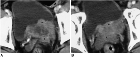
Fig. 1 A 56-year-old male with stage-D1 SCC of the urinary bladder (Case 2).Post-contrast pelvic CT images (A and B) depict a broad-based polypoid mass (m) with nonenhancing cystic or necrotic portions in the left posterior and trigonal area of the urinary bladder. Note the aggressiveness with which this tumor invades seminal vesicles (s), and obliteration of the left ureter.

Fig. 2 A 44-year-old male with stage-C bladder SCC metastasizing to the brain (Case 1).Pre-contrast (A and B) and post-contrast pelvic CT images (C and D) demonstrate a well-enhancing intramural mass in and beyond (arrow) the anterior wall of the urinary bladder and bilateral iliac lymph nodes (n). Another mass in the trigonal area, and multiple distant lymph nodes, are not shown here. Postoperative follow-up brain CT (E) revealed the presence of multiple metastatic nodules (arrowheads), and this patient eventually succumbed to this widespread metastasis (stage D2).
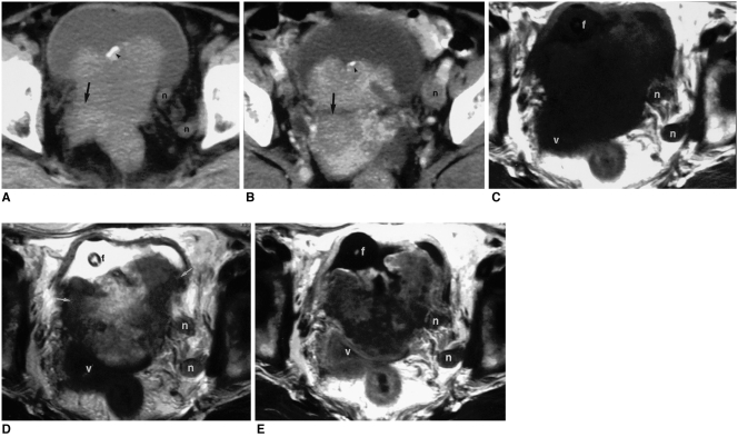
Fig. 3 A 59-year-old female with stage D1 SCC of the urinary bladder (Case 6).A, B. Pre-contrast (A) and post-contrast (B) pelvic CT reveals a heterogeneously enhancing irregular mass in and beyond the posterior wall of the urinary bladder. At the anterior aspect of the mass, there is calcification (arrowhead). Note the exophytic posterior portion of the tumor beyond the bladder wall (arrow), and left pelvic lymphadenopathy (n).C-E. Axial T1-weighted (C), T2-weighted (D), and post-contrast T1-weighted (E) pelvic MR images reveal that the tumor, which invades the anterior vaginal wall (v) and pelvic lymph nodes (n), has both enhancing solid portions and non-enhancing cystic or necrotic portions. A Foley catheter (f) is seen within the bladder. The T2-weighted image shows that the peripheral solid portions of the tumor (arrows) are relatively hypo- or isointense compared with the anterior bladder wall.
Reference
-
1. Cramer SF, Aikawa M, Cebelin M. Neurosecretory granules in small cell invasive carcinoma of the urinary bladder. Cancer. 1981; 47:724–730. PMID: 6261916.
Article2. Fujita K, Nishimura K, Nonomura N, Hirota S, Okuyama A. Early stage small cell carcinoma of the urinary bladder. Int J Urol. 2001; 8:643–644. PMID: 11903694.
Article3. Trias I, Algaba F, Condom E, et al. Small cell carcinoma of the urinary bladder. Presentation of 23 cases and review of 134 published cases. Eur Urol. 2001; 39:85–90. PMID: 11173944.4. Blomjous CEM, Vos W, de Voogt HJD, Valk PVD, Meijer CJLM. Small cell carcinoma of the urinary bladder: a clinicopathologic, morphometric, immunohistochemical, and ultrastructural study of 18 cases. Cancer. 1989; 49:1347–1357. PMID: 2548704.
Article5. Lopez JI, Angulo JC, Flores N, Toledo JD. Small cell carcinoma of the urinary bladder: a clinicopathological study of six cases. Br J Urol. 1994; 49:43–49. PMID: 7507782.
Article6. Holmang S, Borghede G, Johansson SL. Primary small cell carcinoma of the bladder: a report of 25 cases. J Urol. 1995; 153:1820–1822. PMID: 7752326.
Article7. Nejat RJ, Purohit R, Goluboff ET, Petrylak D, Rubin MA, Benson MC. Cure of undifferentiated small cell carcinoma of the urinary bladder with M-VAC chemotherapy. Urol Oncol. 2001; 6:53–55. PMID: 11166621.
Article8. Marshall VF. The relation of the preoperative estimate to the pathologic demonstration of the extent of vesical neoplasm. J Urol. 1952; 68:714. PMID: 12991380.9. Farhat A, Francisco C, Pasquale B, Soloway MS. Small cell carcinoma of the bladder and prostate. Urology. 1995; 46:617–630. PMID: 7495110.
Article10. Abbas F, Civantos F, Benedetto P, et al. Small cell carcinoma of the bladder and prostate. Urology. 1995; 46:617–629. PMID: 7495110.
Article11. Grignon DJ, Ro JY, Ayala AG, et al. Small cell carcinoma of the urinary bladder: a clinicopathologic analysis of 22 cases. Cancer. 1992; 49:527–536. PMID: 1309435.
Article12. Oesterling JE, Brendler CB, Burgers JK, et al. Advanced small cell carcinoma of the bladder. Successful treatment with combined cystoprostatectomy and adjuvant methotrexate, vinblastine, doxorubicin, and cisplatin chemotherapy. Cancer. 1990; 65:1928–1936. PMID: 2164873.
- Full Text Links
-
- Actions
-
Cited
- CITED
-
- Close
- Share
-
- Similar articles
-
- Ultrasound, CT, and MR imaging Findings of Paraganglioma Originating at the Urinary Bladder: A Case Report
- Notice of Redundant Publication
- US, CT and MR Imaging Findings of Leiomyoma of Urinary Bladder: Case Report
- A Case of Small Cell Carcinoma of the Urinary Bladder
- A Case of Primary Undifferentiated Small Cell Carcinoma of the Urinary Bladder
