MRI of a Subcutaneous Myolipoma in the Ankle: a Case Report
- Affiliations
-
- 1Department of Radiology, Daejeon St. Mary's Hospital, The Catholic University of Korea, Daejeon 301-723, Korea.
- 2Department of Orthopedic Surgery, Daejeon St. Mary's Hospital, The Catholic University of Korea, Daejeon 301-723, Korea.
- 3Department of Pathology, Daejeon St. Mary's Hospital, The Catholic University of Korea, Daejeon 301-723, Korea.
- 4Department of Rehabilitation Medicine, Daejeon St. Mary's Hospital, The Catholic University of Korea, Daejeon 301-723, Korea. ces612@nate.com
- KMID: 1116453
- DOI: http://doi.org/10.3348/kjr.2011.12.5.641
Abstract
- Myolipoma is a rare benign tumor, composed of irregularly admixed adipose tissue and smooth muscle fibers. Few literature studies have described the radiologic appearance of myolipoma, especially in the soft tissue. No MRI findings in subcutaneous myolipoma of an extremity have been reported. Here, we report on the case of a 34-year-old woman with myolipoma in the subcutaneous tissue of the ankle and describe MRI features of the lesion.
Keyword
MeSH Terms
Figure
-
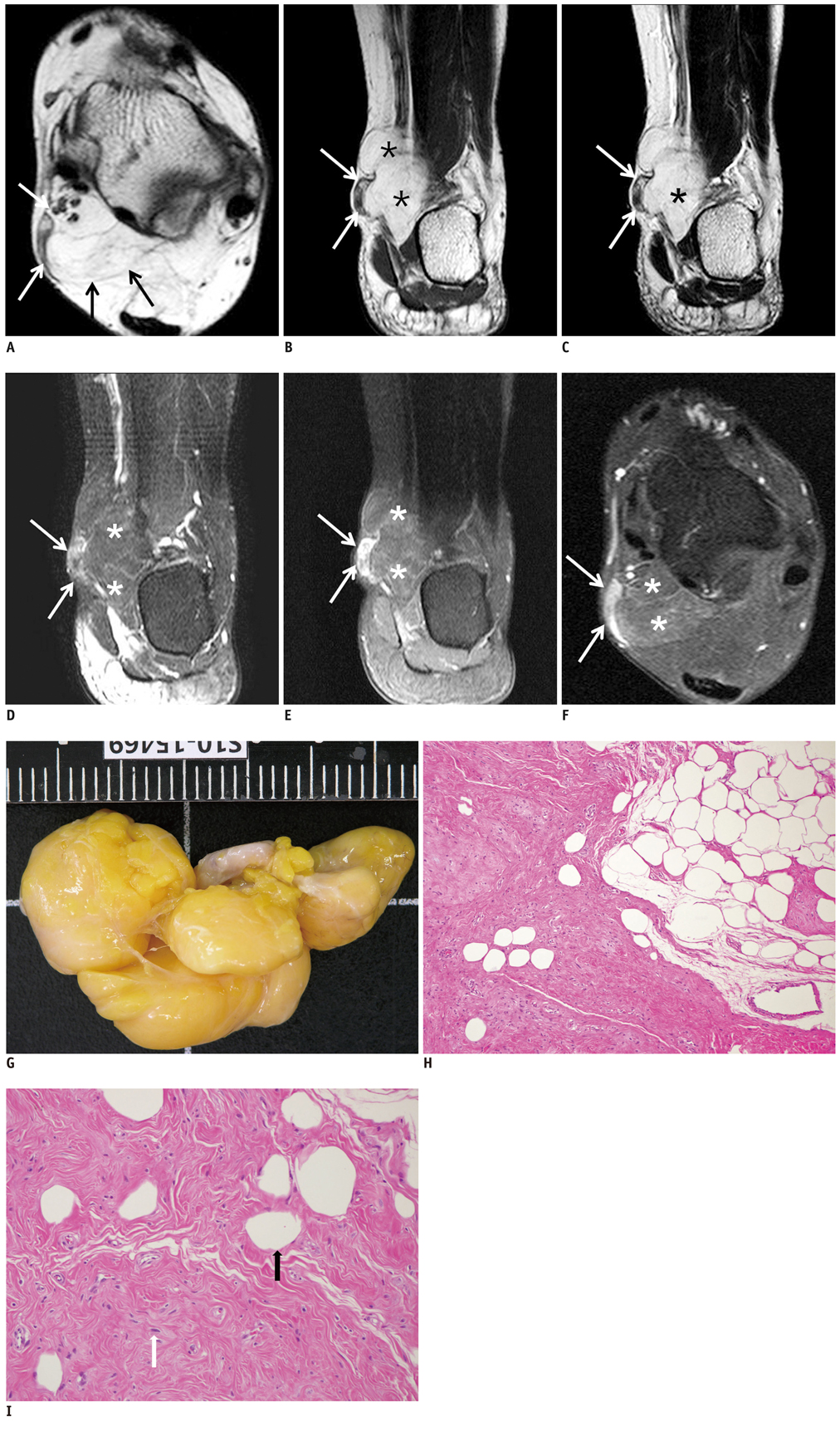
Fig. 1 Myolipoma of subcutaneous region at ankle in 34-year-old woman. A. Axial T1-weighted image shows soft tissue mass (arrows) in subcutaneous region at level of posteromedial ankle joint. B, C. Coronal T1-weighted image (B) and T2-weighted image (C) show that this mass is lobulated in contour with some septations and hyperintense T1 and T2 signal intensity (asterisks). Medial peripheral portion of mass demonstrates intermediate T1 and T2 signal intensity (arrows). D. Coronal fat suppressed T2-weighted image shows significant signal loss of lobulated mass (asterisks) with no fat suppression of peripheral portion of mass (arrows). E, F. Enhanced fat suppressed T1 coronal (E) and axial (F) images demonstrate that main portion of mass shows fat suppression with no remarkable enhancement (asterisks); however, peripheral portion of mass shows heterogeneous enhancement (arrows). G. At gross pathologic examination, tumor is completely encapsulated with yellow to light pink appearance. H, I. Histologic analysis demonstrates variable admixture of smooth muscle (white arrow) and mature adipose tissue (black arrow) (Hematoxylin & Eosin staining; H, × 100; I, × 200).
Reference
-
1. Meis JM, Enzinger FM. Myolipoma of soft tissue. Am J Surg Pathol. 1991. 15:121–125.2. Behranwala KA, Chettiar K, El-Bahrawy M, Stamp G, Kakkar AK. Retroperitoneal myolipoma. World J Surg Oncol. 2005. 3:72.3. Takahashi Y, Imamura T, Irie H, Tanaka F, Fukushima J, Fukusato T, et al. Myolipoma of the retroperitoneum. Pathol Int. 2004. 54:460–463.4. Nagayama A, Miyamura N, Lu Z, Tsuda Y, Kitaoka T, Amemiya T, et al. Light and electron microscopic findings in a patient with orbital myolipoma. Graefes Arch Clin Exp Ophthalmol. 2003. 241:773–776.5. Ben-Izhak O, Elmalach I, Kerner H, Best LA. Pericardial myolipoma: a tumour presenting as a mediastinal mass and containing oestrogen receptors. Histopathology. 1996. 29:184–186.6. Kransdorf MJ, Murphey MD. Imaging of soft tissue tumors. 2006. 2nd ed. Philadelphia: Lippincott Williams & Wilkins;80–149.7. Guillou L, Coindre JM. Newly described adipocytic lesions. Semin Diagn Pathol. 2001. 18:238–249.8. Parratt MT, Gokaraju K, Spiegelberg BG, Miles J, Cannon SR, Briggs TW. Myolipoma affecting the erector spinae: a case report in a child. Case Report Med. 2009. 2009:520126.9. Murphey MD, Carroll JF, Flemming DJ, Pope TL, Gannon FH, Kransdorf MJ. From the archives of the AFIP: benign musculoskeletal lipomatous lesions. Radiographics. 2004. 24:1433–1466.10. Pereira JM, Sirlin CB, Pinto PS, Casola G. CT and MR imaging of extrahepatic fatty masses of the abdomen and pelvis: techniques, diagnosis, differential diagnosis, and pitfalls. Radiographics. 2005. 25:69–85.
