Korean J Radiol.
2011 Oct;12(5):554-558. 10.3348/kjr.2011.12.5.554.
Determination of the rCBF in the Amygdala and Rhinal Cortex Using a FAIR-TrueFISP Sequence
- Affiliations
-
- 1Department of Diagnostic and Interventional Neuro-Radiology, Eberhard-Karls-University, Tubingen, Germany. burkhard.ludescher@med.uni-tuebingen.de
- 2Section on Experimental Radiology, Department of Diagnostic and Interventional Radiology, Eberhard-Karls-University, Tubingen, Germany.
- KMID: 1116440
- DOI: http://doi.org/10.3348/kjr.2011.12.5.554
Abstract
OBJECTIVE
Brain perfusion can be assessed non-invasively by modern arterial spin labeling MRI. The FAIR (flow-sensitive alternating inversion recovery)-TrueFISP (true fast imaging in steady precession) technique was applied for regional assessment of cerebral blood flow in brain areas close to the skull base, since this approach provides low sensitivity to magnetic susceptibility effects. The investigation of the rhinal cortex and the amygdala is a potentially important feature for the diagnosis and research on dementia in its early stages.
MATERIALS AND METHODS
Twenty-three subjects with no structural or psychological impairment were investigated. FAIR-True-FISP quantitative perfusion data were evaluated in the amygdala on both sides and in the pons. A preparation of the radiofrequency FOCI (frequency offset corrected inversion) pulse was used for slice selective inversion. After a time delay of 1.2 sec, data acquisition began. Imaging slice thickness was 5 mm and inversion slab thickness for slice selective inversion was 12.5 mm. Image matrix size for perfusion images was 64 x 64 with a field of view of 256 x 256 mm, resulting in a spatial resolution of 4 x 4 x 5 mm. Repetition time was 4.8 ms; echo time was 2.4 ms. Acquisition time for the 50 sets of FAIR images was 6:56 min. Data were compared with perfusion data from the literature.
RESULTS
Perfusion values in the right amygdala, left amygdala and pons were 65.2 (+/- 18.2) mL/100 g/minute, 64.6 (+/- 21.0) mL/100 g/minute, and 74.4 (+/- 19.3) mL/100 g/minute, respectively. These values were higher than formerly published data using continuous arterial spin labeling but similar to 15O-PET (oxygen-15 positron emission tomography) data.
CONCLUSION
The FAIR-TrueFISP approach is feasible for the quantitative assessment of perfusion in the amygdala. Data are comparable with formerly published data from the literature. The applied technique provided excellent image quality, even for brain regions located at the skull base in the vicinity of marked susceptibility steps.
Keyword
MeSH Terms
Figure
-
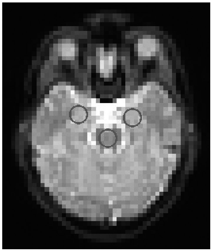
Fig. 1 Region of interest-positions. Positions of region of interests in axial slice.
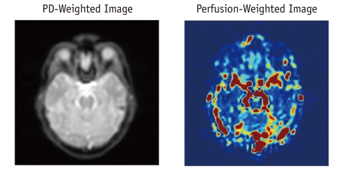
Fig. 2 Proton density (PD) weighted and perfusion weighted images.
Reference
-
2. Cha S. Perfusion MR imaging of brain tumors. Top Magn Reson Imaging. 2004; 15:279–289. PMID: 15627003.
Article3. Chaves CJ, Silver B, Schlaug G, Dashe J, Caplan LR, Warach S. Diffusion- and perfusion-weighted MRI patterns in borderzone infarcts. Stroke. 2000; 31:1090–1096. PMID: 10797170.
Article4. Wintermark M, Sesay M, Barbier E, Borbely K, Dillon WP, Eastwood JD, et al. Comparative overview of brain perfusion imaging techniques. J Neuroradiol. 2005; 32:294–314. PMID: 16424829.
Article5. Choi S, Liu H, Shin TB, Lee JH, Yoon SK, Oh JY, et al. Perfusion imaging of the brain using Z-score and dynamic images obtained by subtracting images from before and after contrast injection. Korean J Radiol. 2004; 5:143–148. PMID: 15467410.
Article6. Kim SG. Quantification of relative cerebral blood flow change by flow-sensitive alternating inversion recovery (FAIR) technique: application to functional mapping. Magn Reson Med. 1995; 34:293–301. PMID: 7500865.
Article7. Kim SG, Tsekos NV. Perfusion imaging by a flow-sensitive alternating inversion recovery (FAIR) technique: application to functional brain imaging. Magn Reson Med. 1997; 37:425–435. PMID: 9055234.
Article8. Calamante F, Thomas DL, Pell GS, Wiersma J, Turner R. Measuring cerebral blood flow using magnetic resonance imaging techniques. J Cereb Blood Flow Metab. 1999; 19:701–735. PMID: 10413026.
Article9. Boss A, Martirosian P, Klose U, Nagele T, Claussen CD, Schick F. FAIR-TrueFISP imaging of cerebral perfusion in areas of high magnetic susceptibility differences at 1.5 and 3 Tesla. J Magn Reson Imaging. 2007; 25:924–931. PMID: 17410577.
Article10. Dai W, Lopez OL, Carmichael OT, Becker JT, Kuller LH, Gach HM. Mild cognitive impairment and alzheimer disease: patterns of altered cerebral blood flow at MR imaging. Radiology. 2009; 250:856–866. PMID: 19164119.
Article11. Martirosian P, Klose U, Mader I, Schick F. FAIR true-FISP perfusion imaging of the kidneys. Magn Reson Med. 2004; 51:353–361. PMID: 14755661.
Article12. Herscovitch P, Raichle ME. What is the correct value for the brain--blood partition coefficient for water? J Cereb Blood Flow Metab. 1985; 5:65–69. PMID: 3871783.
Article13. Devanand DP, Michaels-Marston KS, Liu X, Pelton GH, Padilla M, Marder K, et al. Olfactory deficits in patients with mild cognitive impairment predict Alzheimer's disease at follow-up. Am J Psychiatry. 2000; 157:1399–1405. PMID: 10964854.
Article14. Lee C, Lopez OL, Becker JT, Raji C, Dai W, Kuller LH, et al. Imaging cerebral blood flow in the cognitively normal aging brain with arterial spin labeling: implications for imaging of neurodegenerative disease. J Neuroimaging. 2009; 19:344–352. PMID: 19292827.
Article15. Giovacchini G, Bonwetsch R, Herscovitch P, Carson RE, Theodore WH. Cerebral blood flow in temporal lobe epilepsy: a partial volume correction study. Eur J Nucl Med Mol Imaging. 2007; 34:2066–2072. PMID: 17768621.
Article
- Full Text Links
-
- Actions
-
Cited
- CITED
-
- Close
- Share
-
- Similar articles
-
- MR Imaging of Advanced Gastric Cancer: Comparison between T1-weighted FLASH, T2-weighted TSE, and TrueFISP
- An Experimental Study on the Effects of Bilateral Amygdaloid Destruction
- Differences in Regional Cerebral Blood Flow between Mixed Dementia and Alzheimer’s Disease
- Decreased rCBF in Dpressed Elderly patient with Cognitive Impairment
- The Effect of Nimodipine on Size of Infarction and the Regional Cerebral Blood Flow in Focal Cerebral Ischemia of Rats
