Korean J Radiol.
2011 Oct;12(5):547-553. 10.3348/kjr.2011.12.5.547.
Prevalence of Unruptured Intracranial Aneurysm on MR Angiography
- Affiliations
-
- 1Department of Radiology, Samsung Medical Center, Sungkyunkwan University School of Medicine, Seoul 135-710, Korea. drpjeon@gmail.com
- 2Department of Radiology, National Health Insurance Corporation Ilsan Hospital, Gyeonggi-do 410-719, Korea.
- KMID: 1116439
- DOI: http://doi.org/10.3348/kjr.2011.12.5.547
Abstract
OBJECTIVE
To evaluate the prevalence of incidentally found unruptured intracranial aneurysms (UIAs) on the brain MR angiography (MRA) from a community-based general hospital.
MATERIALS AND METHODS
This was a prospectively collected retrospective study, carried out from January 2004 to December 2004. The subjects included 3049 persons from a community-based hospital in whom MRA was performed according to a standardized protocol in an outpatient setting. Age- and sex-specific prevalence of UIAs was calculated. The results by MRA were compared with intra-arterial digital subtraction angiography (DSA) findings.
RESULTS
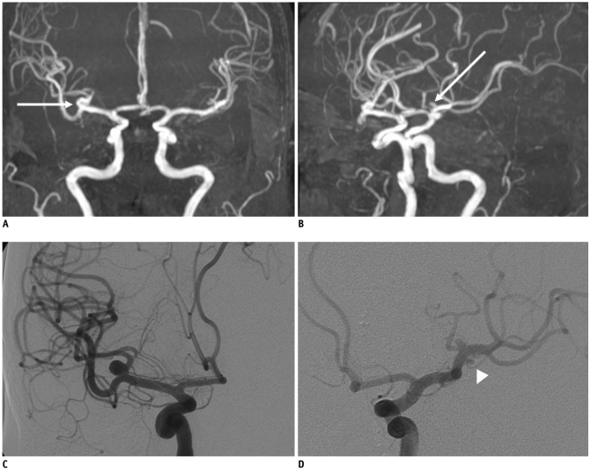
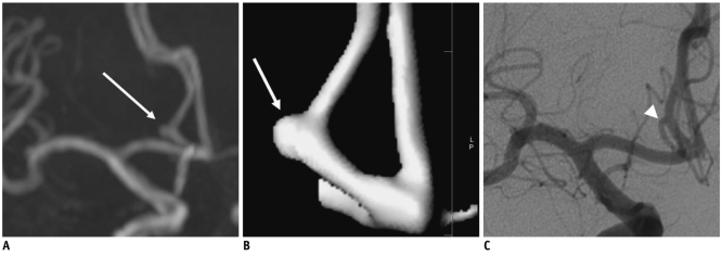
Unruptured intracranial aneurysms were found in 137 (5%) of the 3049 patients (M:F = 43:94; mean age, 60.2 years). The prevalence of UIAs was 5% (n = 94) in women and 4% (n = 43) in men, respectively (p = 0.2046) and showed no age-related increase. The most common site of aneurysm was at the distal internal carotid artery (n = 64, 39%), followed by the middle cerebral artery (n = 40, 24%). In total, 99% of aneurysms measured less than 12 mm, and 93% of aneurysms measured less than 7 mm. Direct comparisons between MRA and DSA were available in 70 patients with 83 UIAs; the results revealed two false positive and two false negative results.
CONCLUSION
This community-hospital based study suggested a higher prevalence of UIAs observed by MRA than previously reported. These findings should be anticipated in the design and use of neuroimaging in clinical practice.
Keyword
MeSH Terms
Figure
Cited by 3 articles
-
Isolated Lenticulostriate Artery Aneurysm Rupture in a Patient with Behçet's Disease
Seongjun Hwang, Sung Hae Chang, Sang Wan Chung, You-Jung Ha, Eun Ha Kang, Yeong Wook Song, Yun Jong Lee
J Rheum Dis. 2015;22(5):317-321. doi: 10.4078/jrd.2015.22.5.317.Prevalence of Unruptured Intracranial Aneurysms: A Single Center Experience Using 3T Brain MR Angiography
Jae Ho Kim, Kyung-Yul Lee, Sang Woo Ha, Sang Hyun Suh
Neurointervention. 2021;16(2):117-121. doi: 10.5469/neuroint.2021.00024.Unruptured Intracranial Aneurysm: Screening, Prevalence and Risk Factors
Bum-soo Kim
Neurointervention. 2021;16(3):201-203. doi: 10.5469/neuroint.2021.00451.
Reference
-
1. Stehbens WE. Aneurysms and anatomical variation of cerebral arteries. Arch Pathol. 1963; 75:45–64. PMID: 14087271.2. Stehbens WE. Pathology of the cerebral blood vessels. 1972. St. Louis: C.V. Mosby.3. Rinkel GJ, Djibuti M, Algra A, van Gijn J. Prevalence and risk of rupture of intracranial aneurysms: a systematic review. Stroke. 1998; 29:251–256. PMID: 9445359.4. Housepian EM, Pool JL. A systematic analysis of intracranial aneurysms from the autopsy file of the Presbyterian Hospital, 1914 to 1956. J Neuropathol Exp Neurol. 1958; 17:409–423. PMID: 13564252.5. Chason JL, Hindman WM. Berry aneurysms of the circle of Willis; results of a planned autopsy study. Neurology. 1958; 8:41–44. PMID: 13493689.
Article6. Iwamoto H, Kiyohara Y, Fujishima M, Kato I, Nakayama K, Sueishi K, et al. Prevalence of intracranial saccular aneurysms in a Japanese community based on a consecutive autopsy series during a 30-year observation period. The Hisayama study. Stroke. 1999; 30:1390–1395. PMID: 10390312.7. Rocque BG, Baskaya MK, Kuo JS. Incidental findings on brain MRI. N Engl J Med. 2008; 358:853. author reply 854-855. PMID: 18287610.
Article8. Horikoshi T, Akiyama I, Yamagata Z, Nukui H. Retrospective analysis of the prevalence of asymptomatic cerebral aneurysm in 4518 patients undergoing magnetic resonance angiography--when does cerebral aneurysm develop? Neurol Med Chir (Tokyo). 2002; 42:105–112. discussion 113. PMID: 11936051.9. Ross JS, Masaryk TJ, Modic MT, Ruggieri PM, Haacke EM, Selman WR. Intracranial aneurysms: evaluation by MR angiography. AJNR Am J Neuroradiol. 1990; 11:449–455. PMID: 2112306.
Article10. White PM, Wardlaw JM, Easton V. Can noninvasive imaging accurately depict intracranial aneurysms? A systematic review. Radiology. 2000; 217:361–370. PMID: 11058629.
Article11. Chung TS, Joo JY, Lee SK, Chien D, Laub G. Evaluation of cerebral aneurysms with high-resolution MR angiography using a section-interpolation technique: correlation with digital subtraction angiography. AJNR Am J Neuroradiol. 1999; 20:229–235. PMID: 10094343.12. Grandin CB, Mathurin P, Duprez T, Stroobandt G, Hammer F, Goffette P, et al. Diagnosis of intracranial aneurysms: accuracy of MR angiography at 0.5 T. AJNR Am J Neuroradiol. 1998; 19:245–252. PMID: 9504473.13. Yue NC, Longstreth WT Jr, Elster AD, Jungreis CA, O'Leary DH, Poirier VC. Clinically serious abnormalities found incidentally at MR imaging of the brain: data from the Cardiovascular Health Study. Radiology. 1997; 202:41–46. PMID: 8988190.
Article14. Mallouhi A, Felber S, Chemelli A, Dessl A, Auer A, Schocke M, et al. Detection and characterization of intracranial aneurysms with MR angiography: comparison of volume-rendering and maximum-intensity-projection algorithms. AJR Am J Roentgenol. 2003; 180:55–64. PMID: 12490476.
Article15. Vernooij MW, Ikram MA, Tanghe HL, Vincent AJ, Hofman A, Krestin GP, et al. Incidental findings on brain MRI in the general population. N Engl J Med. 2007; 357:1821–1828. PMID: 17978290.
Article16. Inagawa T, Hirano A. Autopsy study of unruptured incidental intracranial aneurysms. Surg Neurol. 1990; 34:361–365. PMID: 2244298.
Article17. Wiebers DO, Whisnant JP, Huston J 3rd, Meissner I, Brown RD Jr, Piepgras DG, et al. Unruptured intracranial aneurysms: natural history, clinical outcome, and risks of surgical and endovascular treatment. Lancet. 2003; 362:103–110. PMID: 12867109.
Article18. Guglielmi G, Vinuela F, Dion J, Duckwiler G. Electrothrombosis of saccular aneurysms via endovascular approach. Part 2: Preliminary clinical experience. J Neurosurg. 1991; 75:8–14. PMID: 2045924.19. Russell SM, Lin K, Hahn SA, Jafar JJ. Smaller cerebral aneurysms producing more extensive subarachnoid hemorrhage following rupture: a radiological investigation and discussion of theoretical determinants. J Neurosurg. 2003; 99:248–253. PMID: 12924696.
Article20. Hwang JH, Roh HG, Chun YI, Kang HS, Choi JW, Moon WJ, et al. Endovascular coil embolization of very small intracranial aneurysms. Neuroradiology. 2011; 53:349–357. PMID: 20574735.
Article21. van Rooij WJ, Keeren GJ, Peluso JP, Sluzewski M. Clinical and angiographic results of coiling of 196 very small (< or = 3 mm) intracranial aneurysms. AJNR Am J Neuroradiol. 2009; 30:835–839. PMID: 19131407.22. Gupta V, Chugh M, Jha AN, Walia BS, Vaishya S. Coil embolization of very small (2 mm or smaller) berry aneurysms: feasibility and technical issues. AJNR Am J Neuroradiol. 2009; 30:308–314. PMID: 19001535.
Article23. Inagawa T, Hada H, Katoh Y. Unruptured intracranial aneurysms in elderly patients. Surg Neurol. 1992; 38:364–370. PMID: 1485213.
Article24. Chae KS, Jeon P, Kim KH, Kim ST, Kim HJ, Byun HS. Endovascular coil embolization of very small intracranial aneurysms. Korean J Radiol. 2010; 11:536–541. PMID: 20808697.
Article25. Okahara M, Kiyosue H, Yamashita M, Nagatomi H, Hata H, Saginoya T, et al. Diagnostic accuracy of magnetic resonance angiography for cerebral aneurysms in correlation with 3D-digital subtraction angiographic images: a study of 133 aneurysms. Stroke. 2002; 33:1803–1808. PMID: 12105357.


