Renal Artery Embolization of Perirenal Hematoma in Hemorrhagic Fever with Renal Syndrome: A Case Report
- Affiliations
-
- 1Department of Radiology, Dongguk University International Hospital, Dongguk University College of Medicine, Goyang-si, Korea.
- 2Department of Radiology, Ulsan University Hospital, University of Ulsan College of Medicine, Ulsan-si, Korea.
- 3Department of Internal Medicine, Dongguk University International Hospital, Dongguk University College of Medicine, Goyang-si, Korea.
- KMID: 1110733
- DOI: http://doi.org/10.3348/kjr.2007.8.4.348
Abstract
- Hemorrhagic fever with renal syndrome (HFRS) is an acute viral disease characterized by fever, hemorrhage and renal failure. Among the various hemorrhagic complications of HFRS, spontaneous rupture of the kidney and perirenal hematoma are very rare findings. We report here on a case of HFRS complicated by massive perirenal hematoma, and this was treated with transcatheter arterial embolization.
MeSH Terms
Figure
-
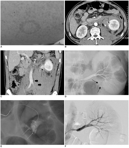
Fig. 1 A 48-year-old man with hemorrhagic fever with renal syndrome complicated by perirenal hematoma. A. Physical examination on admission reveals petechial rashes on the whole body. B. The contrast enhanced CT scan obtained on admission reveals active extravasation of contrast media (arrow) in the inferior portion of the left kidney and perirenal hematoma. C. The coronal reformatted image also shows active extravasation of contrast media at the inferior portion of the left kidney (arrow) with massive perirenal hematoma extending into the pelvic cavity (short arrows). D,E. Selective left renal arteriography (D) and superselective renal arteriography (E) show active extravasation of contrast media from ruptured branches of the inferior division of the renal artery (arrows). There is no hypervascular mass or arteriovenous malformation. F. Selective left renal arteriography performed after superselective embolization with microcoils demonstrates no more extravasation of contrast media.
Cited by 1 articles
-
Reversible Splenium Lesion of the Corpus Callosum in Hemorrhagic Fever with Renal Failure Syndrome
Shin-Hye Baek, Dong-Ick Shin, Hyung-Suk Lee, Sung-Hyun Lee, Hye-Young Kim, Kyeong Seob Shin, Seung Young Lee, Ho-Seong Han, Hyun Jeong Han, Sang-Soo Lee
J Korean Med Sci. 2010;25(8):1244-1246. doi: 10.3346/jkms.2010.25.8.1244.
Reference
-
1. Bruno P, Hassel LH, Brown J, Tanner W, Lau A. The protean manifestations of hemorrhagic fever with renal syndrome. A retrospective review of 26 cases from Korea. Ann Intern Med. 1990. 113:385–391.2. Peters CJ, Simpson GL, Levy H. Spectrum of hantavirus infection: hemorrhagic fever with renal syndrome and hantavirus pulmonary syndrome. Annu Rev Med. 1999. 50:531–545.3. Kim SH, Kim S, Lee JS, Choi BI, Kim YG, Han JS, et al. Hemorrhagic fever with renal syndrome: MR imaging of the kidney. Radiology. 1990. 175:823–825.4. Cho BY, Koh CS, Lee MH. A clinical observations of complications in Korean hemorrhagic fever. Korean J Intern Med. 1979. 22:22–29.5. Guang MY, Liu GZ, Cosgriff TM. Hemorrhage in hemorrhagic fever with renal syndrome in China. Rev Infect Dis. 1989. 11:S884–S890.6. Lee MH, Kim BK, Kim SG, Park SY, Han JS, Kim ST, et al. Coagulopathy in hemorrhagic fever with renal syndrome (Korean hemorrhagic fever). Rev Infect Dis. 1989. 11:S877–S883.7. Valiakhmetov RZ, Gafarov AI, Memkhes VS, Galimzianov VZ. Spontaneous rupture of the kidneys in hemorrhagic fever with renal syndrome. Urol Nefrol (Mosk). 1990. 6:50–53.8. Belville JS, Morgentaler A, Loughlin KR, Tumeh SS. Spontaneous perinephric and subcapsular renal hemorrhage: evaluation with CT, US, and angiography. Radiology. 1989. 172:733–738.9. Nussbaum A, Hunter TB, Stables DP. Spontaneous cyst rupture on renal CT. AJR Am J Roentgenol. 1984. 142:751–752.
- Full Text Links
-
- Actions
-
Cited
- CITED
-
- Close
- Share
-
- Similar articles
-
- A Case of Massive Perirenal Hematoma in a Patient with Hemorrhagic Fever with Renal Syndrome
- A case of retroperitoneal hematoma by spontaneous rupture of renal capsule in hemorrhagic fever with renal syndrome presented with anuria and unilateral flank pain
- A case of ruptured renal cortical arteriovenous malformation of the right testicular vein in hemorrhagic fever with renal syndrome
- A Renal Perforating Artery Mistaken for Arterial Bleeding after Percutaneous Renal Biopsy: A Case Report
- A Case of Hemorrhagic Fever with Renal Syndrome Complicated by Retroperitoneal Hematoma and Hemothorax
