Korean J Ophthalmol.
2008 Mar;22(1):53-57. 10.3341/kjo.2008.22.1.53.
A Case Report on the Change of the Refractive Power After a Blunt Trauma
- Affiliations
-
- 1Department of Ophthalmology, Eulji University School of Medicine, Eulji Medical Center, Seoul, Korea. parkse@eulji.ac.kr
- KMID: 1107492
- DOI: http://doi.org/10.3341/kjo.2008.22.1.53
Abstract
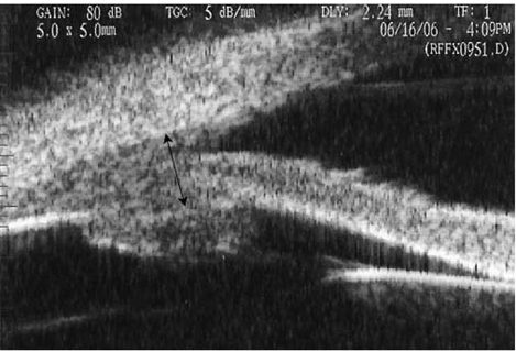
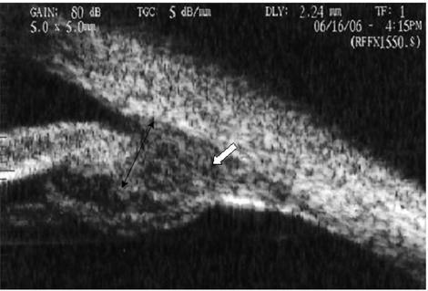
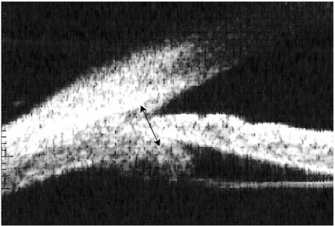
- PURPOSE: To determine the pathogenesis of transient myopia after blunt eye trauma. METHODS: In one patient, the refraction of both eyes (the left eye was injured, but the right eye was not) was measured with an autorefractometer. The cycloplegic refraction was measured at the early stage of trauma and again 3 months after the blunt eye injury. The angle and depth of the anterior chamber, the ciliary body, and the choroids were examined by ultrasound biomicroscopy (UBM) over 3 months. The depth of the anterior chamber, the thickness of the lens, and the axial length were measured by A-scan ultrasonography in both eyes. During the 3 months after the injury, we made comparisons between the menifest and the cycloplegic refractions, the depths of anterior chambers, the thickness of the lenses, the axial lengths, and the UBM-determined appearances of the angles and depths of the anterior chambers, the ciliary bodies, and the choroids in both eyes. RESULTS: We suspect that the depth reduction in the anterior chamber, the increase in anterior to posterior lens diameter, and the edema in the ciliary body are all related to the change in the refractive power following the blunt trauma. CONCLUSIONS: Ultrasound biomicroscopy (UBM) and ultrasonography of the anterior segment in the eye may be helpful to diagnose and confirm changes in the refractive power that occur after trauma.
Keyword
MeSH Terms
Figure
Reference
-
1. Duke Elder S. System of Ophthalmology. 1970. Vol. 5. St.Louis: Mosby;354–355.2. Dotan S, Oliver M. Shallow anterior chamber and uveal effusion after nonperforating trauma to the eye. Am J Ophthalmol. 1982. 94:782–784.3. Kutner BN. Acute angle closure glaucoma in nonperforating blunt trauma. Arch Ophthalmol. 1988. 106:19–20.4. Steele CA, Tullo AB, Marsh IB, Storey JK. Traumatic myopia; an ultrasonographic and clinical study. Br J Ophthalmol. 1987. 71:301–303.5. Romem M, Isakow I, Dolev Z. Posttraumatic transient glaucoma and myopia. Am J Ophthalmol. 1985. 99:495.6. Ikeda N, Ikeda T, Nagata M, Miura O. Pathogenesis of transient high myopia after blunt trauma. Ophthalmology. 2002. 109:501–507.7. Galin MA, Baras I, Zweifach P. Diamox induced myopia. Am J Ophthalmol. 1962. 54:237–240.8. Howes EL Jr, Mckay DG. Circulating immune complexes. Effects on ocular vascular permeability in the rabbit. Arch Ophthalmol. 1975. 93:365–370.9. Fleming DG, Hall JL. Autonomic innervation to ciliary body. Am J Ophthalmol. 1959. 48:287–293.10. Shea M, Mednick EB. Ciliary body reattachment in ocular hypotony. Arch Ophthalmol. 1981. 99:278–281.11. Barasch K, Galin MA, Baras I. Postcyclodialysis hypotony. Am J Ophthalmol. 1969. 68:644–645.12. Chandler PA, Maumenee AE. Major cause of hypotony. Trans Am Acad Ophthalmol Otolaryngol. 1961. 65:563–575.13. Shaffer RN, Weiss DI. Concerning cyclodialysis and hypotony. Arch Ophthalmol. 1962. 68:25–31.14. Brubaker RF, Pederson JE. Ciliochoroidal detachment. Surv Ophthalmol. 1983. 27:281–289.15. Kuchle M, Naumann GO. Direct cyclopexy for traumatic cyclodialysis with persisting hypotony. Ophthalmology. 1995. 102:322–323.
- Full Text Links
- Actions
-
Cited
- CITED
-
- Close
- Share
- Similar articles
-
- A Case of Sudden Refractive Change with Intraocular Pressure Change Following Trauma
- The Accuracy of the Orbscan-derived Total Refractive Power after Laser in Situ Keratomileusis
- Intraocular Lens Power Calculation According to the Difference between Anterior and Total Keratometry Using Scheimpflug Imaging
- Duodenal Injury after Blunt Abdominal Trauma: Report of Two Cases
- Clinical Aspects of Transient Myopia after Blunt Eye Trauma


