J Vet Sci.
2008 Jun;9(2):211-213. 10.4142/jvs.2008.9.2.211.
Macrophages, myofibroblasts and mast cells in a rat liver infected with Capillaria hepatica
- Affiliations
-
- 1Department of Veterinary Pathology, College of Veterinary Medicine, Kyungpook National University, Daegu 702-701, Korea. jeongks@knu.ac.kr
- 2College of Veterinary Medicine, Konkuk University, Seoul 143-701, Korea.
- KMID: 1106239
- DOI: http://doi.org/10.4142/jvs.2008.9.2.211
Abstract
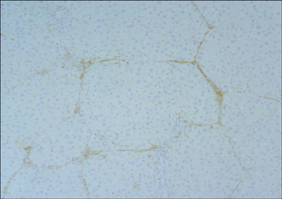
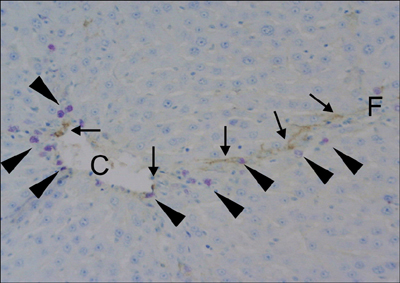
- We trapped a rat (Rattus norvegicus) infected with Capillaria hepatica. At necropsy, grossly yellowish-white nodules (2-3 mm in diameter) were noted to be scattered on the liver's surface. Microscopically, granulomatous and fibrotic nodules that contained the eggs and/or adult worms of Capillaria hepatica were detected in the liver. Septal fibrosis was diffusely formed throughout the liver. There were a number of ED1-positive macrophages located in the sinusoids of the pseudolobules. On the double staining, myofibroblasts and mast cells were generally observed within the fibrous septa with the mast cells in close proximity to the myofibroblasts. We suggest that the interactions between macrophages, myofibroblasts and mast cells play a role in the septal fibrosis observed in rats infected by Capillaria hepatica.
Keyword
MeSH Terms
Figure
Reference
-
1. Akiyoshi H, Terada T. Mast cell, myofibroblast and nerve terminal complexes in carbon tetrachloride-induced cirrhotic rat livers. J Hepatol. 1998. 29:112–119.
Article2. Bhunchet E, Eishi Y, Wake K. Contribution of immune response to the hepatic fibrosis induced by porcine serum. Hepatology. 1996. 23:811–817.
Article3. de Souza MM, Silva LM, Barbosa AA Jr, de Oliveira IR, Parana R, Andrade ZA. Hepatic capillariasis in rats: a new model for testing antifibrotic drugs. Braz J Med Biol Res. 2000. 33:1329–1334.
Article4. Ferreira LA, Andrade ZA. Capillaria hepatica: a cause of septal fibrosis of the liver. Mem Inst Oswaldo Cruz. 1993. 88:441–447.
Article5. Jeong WI, Lee CS, Park SJ, Chung JY, Jeong KS. Kinetics of macrophages, myofibroblasts and mast cells in carbon tetrachloride-induced rat liver cirrhosis. Anticancer Res. 2002. 22:869–877.6. Laskin DL. Non parenchymal cells and hepatotoxicity. Semin Liver Dis. 1990. 10:293–304.7. Li CY, Baek JY. Mastocytosis and fibrosis: role of cytokines. Int Arch Allergy Immunol. 2002. 127:123–126.
Article8. Neafie RC, Connor DH, Cross JH. Binford CH, Connor DH, editors. Capillariasis (intestinal and hepatic). Pathology of Tropical and Extraordinary Diseases. 1976. Vol. 2. Washington DC: Armed Forces Institute of Pathology;481–492.9. Oliveira RF, Andrade ZA. Worm load and septal fibrosis of the liver in Capillaria hepatica-infected rats. Mem Inst Oswaldo Cruz. 2001. 96:1001–1003.
Article10. Ramos SG, Montenegro AP, Goissis G, Rossi MA. Captopril reduces collagen and mast cell and eosinophil accumulation in pig serum-induced rat liver fibrosis. Pathol Int. 1994. 44:655–661.
Article11. Santos AB, Tolentino M Jr, Andrade ZA. Pathogenesis of hepatic septal fibrosis associated with Capillaria hepatica infection of rats. Rev Soc Bras Med Trop. 2001. 34:503–506.
Article12. Shiga A, Shirota K, Ikeda T, Nomura Y. Morphological and immunohistochemical studies on porcine serum-induced rat liver fibrosis. J Vet Med Sci. 1997. 59:159–167.
Article
- Full Text Links
- Actions
-
Cited
- CITED
-
- Close
- Share
- Similar articles
-
- Prevalence of Capillaria hepatica among house rat in Seoul
- Studies on the parasitic helminths of Korea II. Parasites of the rat, Rattus norvegicus Erxl. in Seoul, with the description of Capillaria hepatica(Bancroft, 1893) Travassos, (1915)
- Changes of cytokine mRNA expression and IgG responses in rats infected with Capillaria hepatica
- The First Case of Capillaria hepatica Infection in a Nutria (Myocastor coypus) in Korea
- Fates of degranulated mast cells and extruded granules responded to homologous antigen in rats infected with Clonorchis sinensis


