Association of Cholesterol Granuloma and Aspergillosis in the Sphenoid Sinus
- Affiliations
-
- 1Department of Radiology, Seoul Veterans Hospital, Seoul, Korea. knroo@hanmail.net
- 2Department of Pathology, Seoul Veterans Hospital, Seoul, Korea.
- KMID: 1100100
- DOI: http://doi.org/10.3348/kjr.2008.9.s.s30
Abstract
- Cholesterol granuloma (CG) is usually associated with chronic middle ear disease, and is not common in the paranasal sinuses. Additionally, it is very rare for cases of CG to be associated with a fungal infection. However, in this paper, we report a case of sphenoid sinus CG that is associated with aspergilloma in a 78-year-old male patient who presented with right hemifacial pain, headache and toothache. CT revealed the presence of an expansile cystic mass lesion in the sphenoid sinus that showed a high signal intensity on both the T1 and T2 weighted images. This mass was later determined to be CG. The suspected etiologic mechanisms of both CG and aspergilloma of the paranasal sinuses are similar, and impaired drainage and obstruction of the ventilation of the paranasal sinuses are considered to be the causative mechanism of both diseases. Overall, the results of this study indicate that the use of MRI findings could be helpful for differentiating CG from other paranasal sinus mass lesions.
Keyword
MeSH Terms
Figure
-
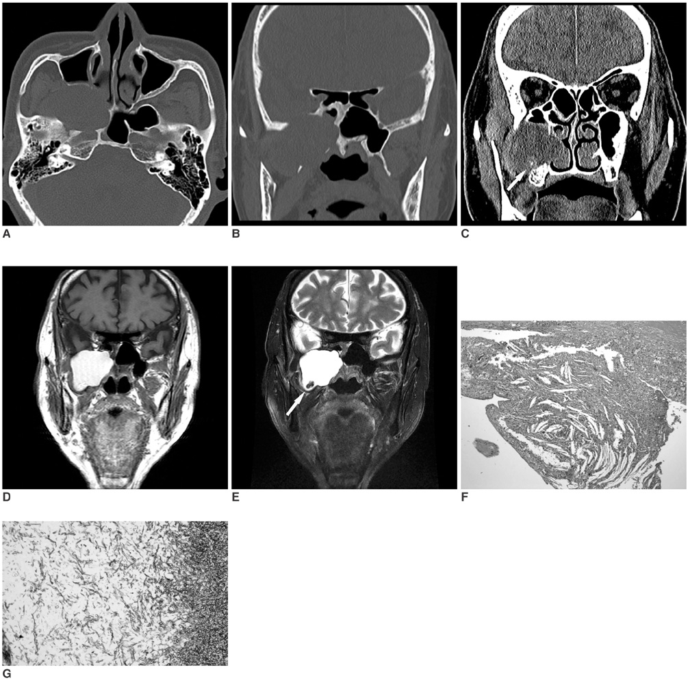
Fig. 1 78-year-old male patient with right infratemporal fossa mass arising from right sphenoid sinus. A-C. Axial CT of sphenoid sinus showing 4.8 × 3.7-cm soft tissue mass lesion arising from right sphenoid sinus (A). Upon coronal CT scan, mass lesion was shown to extend to right infratemporal and pterygopalatine fossae and indent surrounding bony structure. In addition, right sphenoid sinus bony wall thickening and sclerotic change is also seen (B). Coronal CT scan revealed tiny calcified lesion, believed to be fungal ball, at inferior portion of mass (white arrow in C). D, E. Coronal T1WI scan, which shows high signal intensity in mass legion that is located at right infratemporal and pterygopalatine fossae (D). T2WI scan, which reveals mass lesion with high signal intensity and 10 × 7-mm-sized lesion with low signal intensity, which was believed to be fungal ball, at inferior portion of mass (white arrow in E). F. Mass lesion characterized by cholesterol clefts surrounded by giant cells and foamy histiocytes (Hematoxylin & Eosin staining, original magnification ×200). G. Photomicrograph of excisional tumor specimen showing septated hyphae typical of Aspergillus infection (Grocott-Gomori methenamine silver nitrate staining, magnification ×400).
Reference
-
1. Sarioglu S, Pabuççuoglu U, Arzu Topal N. Cholesterol granuloma and aspergilloma of the maxillary sinus. Eur Arch Otorhinolaryngol. 2001. 258:74–76.2. Bella Z, Torkos A, Tiszlavicz L, Iván L, Jóri J. Cholesterol granuloma of the maxillary sinus resembling an invasive, destructive tumor. Eur Arch Otorhinolaryngol. 2005. 262:531–533.3. Ramani P, Murugesan K, Chandrasekar T, Anuja N. Cholesterol granuloma of maxillary sinus. Int J Oral Maxillofac Surg. 2006. 35:1063–1065.4. Ko MT, Hwang CF, Kao YF, Lui CC, Huang CC, Peng JP. Cholesterol granuloma of the maxillary sinus presenting as sinonasal polyp. Am J Otolaryngol. 2006. 27:370–372.5. Weiland DA, Aygun N. An unusual presentation of a cholesterol granuloma in a pneumatized pterygoid process of the sphenoid sinus. Otolaryngol Head Neck Surg. 2007. 136:153–154.6. Ishizaki M, Ohsumi S, Takashima S, Mandai K. Two cases of cholesterol granuloma of the breast. Breast Cancer. 2001. 8:158–161.7. Kikuchi T, So E, Ishimaru K, Miyabe Y, Abe K, Kobayashi T. Endoscopic sinus surgery in cases of cholesterol granuloma of the maxillary sinus. Tohoku J Exp Med. 2002. 197:233–237.8. Gray WC, Salcman M, Rao KC, Hafiz MA. Cholesterol granuloma of the petrous apex and sphenoidal sinus: a case report. Neurosurgery. 1985. 17:67–69.9. Chao TK. Cholesterol granuloma of the maxillary sinus. Eur Arch Otorhinolaryngol. 2006. 263:592–597.10. Eloy JA, Bederson JB, Smouha EE. Petrous apex aspergillosis as a long-term complication of cholesterol granuloma. Laryngoscope. 2007. 117:1199–1201.11. Som PM, Dillon WP, Curtin HD, Fullerton GD, Lidov M. Hypointense paranasal sinus foci: differential diagnosis with MR imaging and relation to CT findings. Radiology. 1990. 176:777–781.12. Zinreich SJ, Kennedy DW, Malat J, Curtin HD, Epstein JI, Huff LC, et al. Fungal sinusitis: diagnosis with CT and MR imaging. Radiology. 1988. 169:439–444.
