Leiomyosarcoma of the Ovarian Vein: a Case Report with Radiological Findings
- Affiliations
-
- 1Department of Diagnostic Radiology, Inje University College of Medicine, Seoul Paik Hospital, Seoul, Korea. meditererian@hanmail.net
- 2Department of General Surgery, Inje University College of Medicine, Seoul Paik Hospital, Seoul, Korea.
- 3Department of Pathology, Inje University College of Medicine, Seoul Paik Hospital, Seoul, Korea.
- KMID: 1100096
- DOI: http://doi.org/10.3348/kjr.2008.9.s.s14
Abstract
- Leiomyosarcomas of the ovarian vein are very rare. Four cases have been reported in the English language clinical literature. We present a case of leiomyosarcomas where the use of multi-detector CT had a substantial role in the establishment of the preoperative diagnosis. The radiological images as well as intraoperative features are illustrated. We also discuss the radiological findings of the ovarian vein leiomyosarcoma in comparison with those of other venous or retroperitoneal leiomyosarcomas. We expect that the use of multi-detector CT will be the choice for the diagnostic work-up of vascular leiomyosarcomas.
MeSH Terms
Figure
-
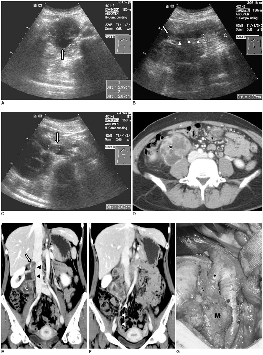
Fig. 1 Radiological and intraoperative findings of ovarian vein leiomyosarcoma in 39-year-old woman. A. Transverse sonography reveals well-defined, spherical, medium-echoic and solid mass with eccentric cystic portion (arrow) at right paravertebral region. B, C. Longitudinal and transverse sonography shows vascular structure, filled with solid components (arrowheads) and connected to main mass (circle), of which superior end shows round protrusion (arrows) in lumen of junctional area of inferior vena cava and right renal vein. D. Just below level of aortic bifurcation, axial contrast-enhanced CT scan shows lobulated and well-defined lobulated retroperitoneal mass with highly enhanced solid components and extensive cystic degenerations (asterisk) at right paravertebral region. E, F. Coronal contrast-enhanced CT scans show vertical pedicular solid structures (black arrowheads), connected to upper portion of mass (circle), which protrudes in lumen of junctional area of right renal vein and inferior vena cava (arrow). Inferior portion of mass connects with normal ovarian vein in pelvis (white arrowheads). G. During surgery, mass (M) is contiguous with right ovarian vein. Note distended right ovarian vein with tumor thrombi (asterisk). En bloc resection of mass together with right kidney, right ureter and retroperitoneal tissue, was performed.
Reference
-
1. Kevorkian J, Cento CP. Leiomyosarcoma of large arteries and veins. Surgery. 1973. 73:390–400.2. Dzsinich C, Gloviczki P, van Heerden JA, Nagorney DM, Pairolero PC, Johnson CM, et al. Primary venous leiomyosarcoma: a rare but lethal disease. J Vasc Surg. 1992. 15:595–603.3. Honore LH, Robins RE, Taylor RH. Leiomyosarcoma of the right ovarian vein a case report. Angiology. 1977. 28:285–288.4. Kawai K, Horiguchi H, Sekido N, Akaza H, Koiso K. Leiomyosarcoma of the ovarian vein: an unusual cause of severe abdominal and flank pain. Int J Urol. 1996. 3:234–236.5. Iannelli A, Karimdjee BS, Fabiani P, Liolos J, Avallone S, Gugenheim J. Leiomyosarcoma of the ovarian vein: report of a case. Int Surg. 2003. 88:6–8.6. Hartman DS, Hayes WS, Choyke PL, Tibbetts GP. Leiomyosarcoma of the retroperitoneum and inferior vena cava: radiologic-pathologic correlation. Radiographics. 1992. 12:1203–1220.7. Young R, Friedman AC, Hartman DS. Computed tomography of leiomyosarcoma of the inferior vena cava. Radiology. 1982. 145:99–103.8. Singh-Panghaal S, Karcnik TJ, Wachsberg RH, Baker SR. Inferior vena caval leiomyosarcoma: diagnosis and biopsy with color Doppler sonography. J Clin Ultrasound. 1997. 25:275–278.9. Blum U, Wildanger G, Windfuhr M, Laubenberger J, Freudenberg N, Munzar T. Preoperative CT and MR imaging of inferior vena cava Leiomyosarcoma. Eur J Radiol. 1995. 20:23–27.
- Full Text Links
-
- Actions
-
Cited
- CITED
-
- Close
- Share
-
- Similar articles
-
- Leiomyosarcoma of the Femoral Vein Mimicking a Peripheral Nerve Sheath Tumor: A Case Report
- Imaging Findings of Primary Adrenal Leiomyosarcoma: A Case Report
- Leiomyosarcoma arising in the great saphenous vein: a case report
- Unusual presentation of retroperitoneal leiomyosarcoma mimicking an adnexal tumor with highly elevated serum CA-19-9
- Primary Ovarian Leiomyosarcoma: A case report
