Korean J Ophthalmol.
2006 Mar;20(1):1-6. 10.3341/kjo.2006.20.1.1.
Clinical Results of Endoscopic Dacryocystorhinostomy using a Microdebrider
- Affiliations
-
- 1Department of Ophthalmology, Dongkang General Hospital, Ulsan, Korea. lavie2k@korea.com
- KMID: 1099065
- DOI: http://doi.org/10.3341/kjo.2006.20.1.1
Abstract
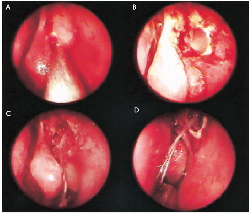
- PURPOSE: The success rate of endoscopic dacryocystorhinostomy has been increasing with the development of better instruments and techniques. We conducted this study to evaluate the clinical results of endoscopic dacryocystorhinostomy using a Microdebrider, which has also been used for functional endoscopic sinus surgery. METHODS: We selected 76 patients (with a total of 84 affected eyes) who had been diagnosed with a nasolacrimal duct obstruction. These patients underwent an endoscopic dacryocystorhinostomy using a Microdebrider, which removed both nasal mucosa and lacrimal sac mucosa and also trimmed the margins of the ostotomy site. We assessed patients' outcomes on an anatomical basis using a dye test and endoscopy, which were used to define the anatomical success. We also arbitrarily defined functional success as whether the subjective epiphora was absent. At a four to 18 month follow-up, we monitored the clinical course to examine any recurrent episodes. RESULTS: The symptoms were alleviated in 72 eyes, with a primary success rate of 85.7%. On nasal endoscopy, a functional failure was seen in four eyes. In these four eyes, the orifice was narrowed by the presence of either granulation tissue or conjunctivochalasis. By contrast, surgical outcomes were the anatomical failure in eight eyes. In these eight eyes, the orifice was obstructed by the presence of granulation tissue as well as the adhesion of nasal mucosa. CONCLUSIONS: Endoscopic dacryocystorhinostomy using a Microdebrider enabled us to make the large fistula while minimizing the damage of adjacent tissue. It might be the recommended surgery that reduces the complications and enhances the success rate.
MeSH Terms
Figure
Reference
-
1. Toti A. Nuovo metodo conservative dicura radicale delle sopourazioni croniche del sacco lacrimale (dacriocistorhinostomia). Clin Moderna. 1904. 10:385–387.2. Caldwell GW. Two new operations of obstruction of the nasal duct with preservation of the canaliculi. Am J Ophthalmol. 1893. 10:189.3. West M. A window resection of the nasal duct in case of stenosis. Trans Am Ophthalmol Soc. 1910. 12:654–658.4. Macbeth R. Problems of lacrimal obstruction: the rhinological approach. Trans Opthal Soc U K. 1956. 76:355–368.5. Lee HC, Chung WS. Success Rate of Endonasal Dacryocystorhinostomy. J Korean Ophthalmol Soc. 1996. 37:211–218.6. Jones LT. Ananatomic approach to problems of the eyelids and lachrymal apparatus. Arch Ophthalmol. 1961. 66:111–124.7. Becker BB. Dacryocystorhinostomy without flaps. Ophthalmic Surg. 1988. 19:419–427.8. Jokinen K, Karja J. Endonasal dacryocystorhinostomy. Arch Otolaryngol. 1974. 100:41–44.9. Berryhill BH, Dorenbusch AA. Twenty years experience with intranasal transseptal dacryocystorhinostomy. Laryngoscope. 1982. 92:379–381.10. McDonogh M, Meiring JH. Endocopic transnasal dacrycystorhinostomy. J Larnygol Otol. 1989. 103:585–587.11. Massaro BM, Gonnering RS, Harris GJ. Endonasal laser dacryocystorhinostomy. A new approach to nasolacrimal duct obstruction. Arch Ophthalmol. 1990. 108:1172–1176.12. Gonnering RS, Lyon DB, Fisher JC. Endoscopic laserassisted lacrimal surgery. Am J Ophthalmol. 1991. 111:152–157.13. Bartley GB. The pros and cons of laser dacryocystorhinostomy. Am J Ophthalmol. 1994. 117:103–106.14. Woog JJ, Metson R, Puliafito CA. Holmium:YAG endonasal laser dacryocystorhinostomy. Am J Ophthalmol. 1993. 116:1–10.15. Kong YT, Kim TI, Kong BW. A report of 131 cases of endoscopic laser lacrimal surgery. Ophthalmology. 1994. 101:1793–1800.16. Javate RM, Campomanes BS, Co ND, et al. The endoscope and the radiofrequency unit in DCR surgery. Ophthal Plast Reconstr Surg. 1995. 11:54–58.17. Hawke WM, McCombe AW. How I do it: nasal polypectomy with an arthroscopic bone shaver: the Stryker "Hummer". J Otolaryngol. 1995. 24:57–59.18. Ferguson BJ, DiBiase PA, D'Amico F. Quantitative analysis of microdebriders used in endoscopic sinus surgery. Am J Otolaryngol. 1999. 20:294–297.19. Unlu HH, Goktan C, Aslan A, Tarhan S. Injury to the lacrimal apparatus after endoscopic sinus surgery: surgical implications from active transport dacryocystography. Otolaryngol Head Neck Surg. 2001. 124:308–312.20. Becker DG. Technical considerations in powered instrumentation. Otolaryngol Clin North Am. 1997. 30:421–434.21. Linberg JV, Anderson RL, Bumsted RM, Barreras R. Study of intranasal ostium external dacryocystorhinostomy. Arch Ophthalmol. 1982. 100:1758–1762.22. Welham RA, Henderson PH. Results of dacryocystorhinostomy analysis of causes for failure. Trans Ophthalmol Soc UK. 1973. 93:601–609.23. Allen KM, Berlin AJ, Levine HL. Intranasal endoscopic analysis of dacrocystorhinostomy failure. Ophthal Plast Reconstr Surg. 1988. 43:143–145.24. Allen KM, Berlin AJ. Dacryocystorhinostomy failure: association with nasolacrimal silicone intubation. Ophthalmic Surg. 1989. 20:486–489.25. Lee TS, Kim SW. The effects of placement of bicanalicular silicone tube and silicone stent on granuloma formation in endoscopic intranasal dacryocystorhinostomy. J Korean Ophthalmol Soc. 1999. 40:16–22.
- Full Text Links
- Actions
-
Cited
- CITED
-
- Close
- Share
- Similar articles
-
- A Case of Retrobulbar Hemorrhage after Endoscopic Sinus Surgery with Microdebrider
- Injury of the Medial Rectus Muscle by Using a Microdebrider During Endoscopic Sinus Surgery : A Case Report
- Endoscopic Dacryocystorhinostomy with Mitomycin-C Application
- Transoral Endoscopic Adenoidectomy with the Microdebrider
- Endoscopic Laser Dacryocystorhinostomy


