A Solitary Fibrofolliculoma in the Eyelid
- Affiliations
-
- 1Department of Ophthalmology, Dankook University College of Medicine, Cheonan, Korea. cataract@empal.com
- KMID: 1099034
- DOI: http://doi.org/10.3341/kjo.2007.21.3.169
Abstract
- PURPOSE: To report the first case of a solitary eyelid fibrofolliculoma and to review the literature. METHODS: A 37-year-old female patient visited the outpatient department with a lesion in the right upper lid that had been growing steadily for a year. The patient had visited the local clinic, and under the diagnosis of chalazion had received incision and curettage twice, but the lesion had recurred. RESULTS: The 5 x 5 mm lesion was located near the upper lid margin. It was a red, hemispheric, smooth nodule, relatively solid to palpation and not painful. Excision and biopsy were performed, and through a histological exam, the diagnosis of fibrofolliculoma was later confirmed. CONCLUSIONS: Solitary fibrofolliculoma is rare, and to the authors' knowledge, a lesion arising in the eyelid has not yet been reported. Fibrofolliculoma should be included in the differential diagnosis when a localized mass lesion arising in the eyelid is encountered.
Keyword
MeSH Terms
Figure
-
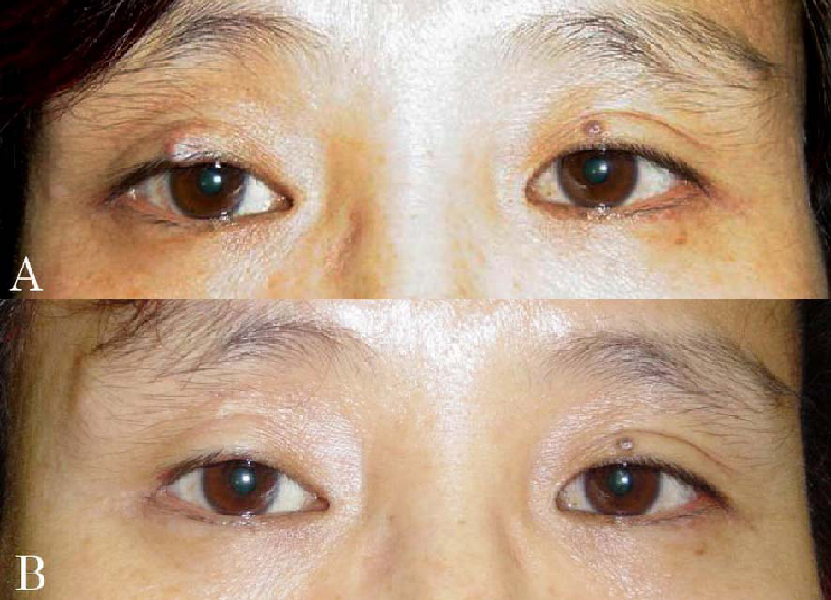
Fig. 1 (A) Preoperative appearance shows a solitary, firm, non-tender, well-demarcated and skin-colored bean-sized mass on the right upper eyelid. (B) Postoperative (11 months) appearance shows the right upper lid, which has maintained its shape; the lesion has not yet recurred.
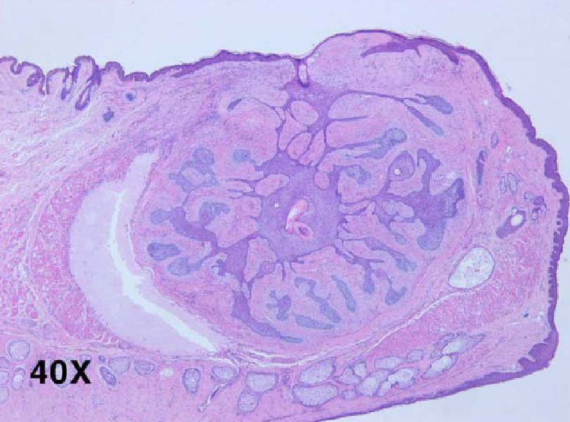
Fig. 2 Low-power histopathologic appearance of a fibrofolliculoma showing proliferation of fibrotic stroma surrounding the central dilated infundibulum of the hair follicle with proliferative thin epithelial strands (Hematoxylin-eosin stain ×40).
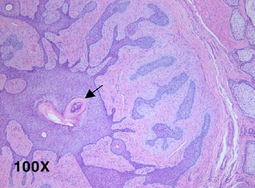
Fig. 3 High-power histopathologic photograph showing the characteristic appearances of proliferating infundibular epithelial strands with perifollicular fibrous reaction, anastomosing to form an epithelial network (arrow: hair follicle, Hematoxylin-eosin stain, ×100).
Reference
-
1. Birt AR, Hogg GR, Dubé WJ. Hereditary multiple fibrofolliculomas with trichodiscomas and acrochordons. Arch Dermatol. 1977. 113:1674–1677.2. Weintraub R, Pinkus H. Multiple fibrofolliculomas (Birt-Hogg-Dubé) associated with a large connective tissue nevus. J Cutan Pathol. 1977. 4:289–299.3. Scully K, Bargman H, Assaad D. Solitary fibrofolliculoma. J Am Acad Dermatol. 1984. 11:361–363.4. Fujita WH, Barr RJ, Headley JL. Multiple fibrofolliculomas with trichodiscomas and acrochordons. Arch Dermatol. 1981. 117:32–35.5. Pinkus H, Coskey R, Burgess GH. Trichodiscoma. A benign tumor related to haarscheibe (hair disk). J Invest Dermatol. 1974. 63:212–218.6. Starink TM, Brownstein MH. Fibrofolliculoma: solitary and multiple types. J Am Acad Dermatol. 1987. 17:493–496.7. Foucar K, Rosen T, Foucar E, et al. Fibrofolliculoma: a clinicopathologic study. Cutis. 1981. 28:429–432.8. Rahbari H, Mehregan AH. Benign follicular neoplasias. J Dermatol Surg Oncol. 1979. 5:295–298.9. Starink TM, Kisch LS, Meijer CJ. Familial multiple trichodiscomas. A clinicopathologic study. Arch Dermatol. 1985. 121:888–891.10. Mo HJ, Park CJ, Yi JY. A Case of Solitary Fibrofolliculoma. Korean J Dermatol. 2001. 39:602–604.11. Kim KH, Choi JH, Lee YS. A Case of Solitary Fibrofolliculoma. Korean J Dermatol. 1984. 22:672–674.12. Lee HK, Maeng LS, Kang SJ, et al. Solitary fibrofolliculoma: A case report. Kor J Pathol. 1996. 30:460–462.
