Grading Anterior Cruciate Ligament Graft Injury after Ligament Reconstruction Surgery: Diagnostic Efficacy of Oblique Coronal MR Imaging of the Knee
- Affiliations
-
- 1Department of Radiology and Institute of Radiation Medicine, Seoul National University College of Medicine, Seoul, Korea. drhong@snu.ac.kr
- 2Department of Radiology, Konkuk University School of Medicine, Seoul, Korea.
- 3Department of Radiology and Center for Imaging Science, Samsung Medical Center, Sungkyunkwan University School of Medicine, Seoul, Korea.
- KMID: 1098195
- DOI: http://doi.org/10.3348/kjr.2008.9.2.155
Abstract
OBJECTIVE
The purpose of this study was to evaluate the diagnostic efficacy of using additional oblique coronal MRI of the knee for grading anterior cruciate ligament (ACL) graft injury after ligament reconstruction surgery. MATERIALS AND METHODS: We retrospectively reviewed 51 consecutive MR knee examinations of 48 patients who underwent both ACL reconstruction and follow-up arthroscopy. The MR examinations included the orthogonal axial, sagittal, coronal images and the oblique coronal T2-weighted images, which were oriented in parallel with the course of the femoral intercondylar roof. Two radiologists independently evaluated the status of the ACL grafts with using the routine knee MRI and then with adding the oblique coronal imaging. The severity of ACL graft injury was graded using a 3-point system from MR images as intact, partial tear or complete tear, and the results were compared with the arthroscopic results. Weighted kappa statistics were used to analyze the diagnostic accuracies of the knee MRI with and without the additional oblique coronal imaging. For each evaluation, the observers reported a confidence level for grading the ACL graft injuries in the two imaging groups. RESULTS: The weighted kappa values according to the routine knee MRI were 0.555 (reader 1) and 0.515 (reader 2). The inclusion of additional oblique coronal imaging increased the weighted kappa values to 0.666 (reader 1) and 0.611 (reader 2). The mean confidence levels by each reader were significantly higher (p < 0.01, paired t-test) with the additional oblique coronal imaging than by using the routine knee MRI alone. CONCLUSION: The additional use of oblique coronal MRI of the knee improves both the diagnostic accuracy and confidence for grading ACL graft injury.
MeSH Terms
Figure
-

Fig. 1 Oblique coronal MR images of anterior cruciate ligament grafts. A. Sagittal T1-weighted MR image is used to localize oblique coronal imaging planes parallel to femoral intercondylar roof. B. Oblique coronal T2-weighted images (TR/TE, 3500/96) show homogeneously dark, straight ligament, suggesting intact anterior cruciate ligament graft (arrows) along its entire length. Both femoral (black arrowheads) and tibial (white arrowheads) sides are clearly demonstrated. Cross section of posterior cruciate ligament (thin arrow) is also shown.
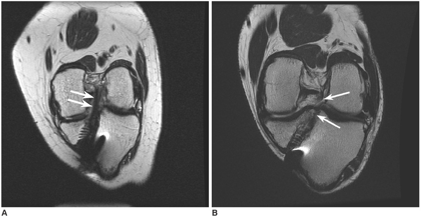
Fig. 2 Grade 1 and 2 injuries in anterior cruciate ligament grafts. A. Oblique coronal T2-weighted image (TR/TR, 4000/96) shows decreased thickness (arrows) of anterior cruciate ligament graft, suggesting grade 1 injury. B. Oblique coronal T2-weighted image (TR/TE, 3000/96) of another patient shows lack of continuity and indistinct contour (arrows) at distal portion of anterior cruciate ligament graft, suggesting grade 2 injury.
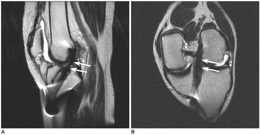
Fig. 3 Grade 0 versus grade 1 anterior cruciate ligament graft injury. A. Sagittal T2-weighted image (TR/TE, 2200/90) shows increased signal intensity and decreased thickness (arrows) of anterior cruciate ligament graft at distal portion, suggesting grade 1 injury. B. Oblique coronal T2-weighted image (TR/TE, 3000/96) shows preserved continuity of anterior cruciate ligament graft with homogeneously dark signal intensity (arrows), suggesting likelihood of grade 0 injury rather than grade one injury. Arthroscopic examination one day later confirmed intact graft with grade 0 injury.
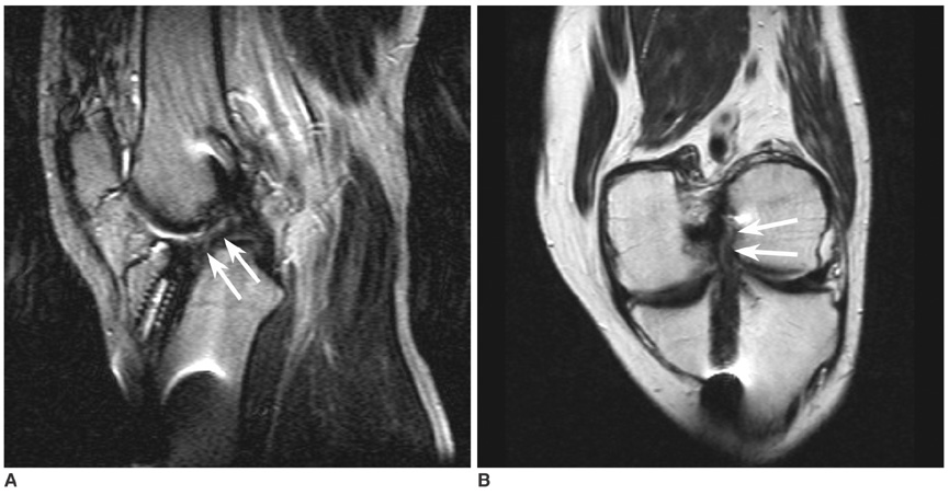
Fig. 4 Grade 1 versus grade 2 anterior cruciate ligament graft injury. A. Sagittal T2-weighted image (TR/TE, 2200/90) shows lack of continuity and indistinct contour (arrows) of anterior cruciate ligament graft, suggesting grade 2 injury. B. Oblique coronal T2-weighted image (TR/TE, 3000/96) shows diffuse thinning, but also partially preserved continuity (arrows), suggesting grade 1 injury. Arthroscopic examination four weeks later confirmed grade 1 injury with graft attenuation with two thirds residual mass.
Reference
-
1. Horton LK, Jacobson JA, Lin J, Hayes CW. MR imaging of anterior cruciate ligament reconstruction graft. AJR Am J Roentgenol. 2000. 175:1091–1097.2. Howell SM, Berns GS, Farley TE. Unimpinged and impinged anterior cruciate ligament grafts: MR signal intensity measurements. Radiology. 1991. 179:639–643.3. Papakonstantinou O, Chung CB, Chanchairujira K, Resnick DL. Complications of anterior cruciate ligament reconstruction: MR imaging. Eur Radiol. 2003. 13:1106–1117.4. Rak KM, Gillogly SD, Schaefer RA, Yakes WF, Liljedahl RR. Anterior cruciate ligament reconstruction: evaluation with MR imaging. Radiology. 1991. 178:553–556.5. Recht MP, Kramer J. MR imaging of the postoperative knee: a pictorial essay. Radiographics. 2002. 22:765–774.6. Schatz JA, Potter HG, Rodeo SA, Hannafin JA, Wickiewicz TL. MR imaging of anterior cruciate ligament reconstruction. AJR Am J Roentgenol. 1997. 169:223–228.7. Hong SJ, Ahn JM, Ahn JH, Park SW. Postoperative MR findings of the healthy ACL grafts: correlation with second look arthroscopy. Clin Imaging. 2005. 29:55–59.8. Min BH, Chung WY, Cho JH. Magnetic resonance imaging of reconstructed anterior cruciate ligament. Clin Orthop Relat Res. 2001. 393:237–243.9. Murakami Y, Sumen Y, Ochi M, Fujimoto E, Adachi N, Ikuta Y. MR evaluation of human anterior cruciate ligament autograft on oblique axial imaging. J Comput Assist Tomogr. 1998. 22:270–275.10. Duc SR, Zanetti M, Kramer J, Kach KP, Zollikofer CL, Wentz KU. Magnetic resonance imaging of anterior cruciate ligament tears: evaluation of standard orthogonal and tailored paracoronal images. Acta Radiol. 2005. 46:729–733.11. Hong SH, Choi JY, Lee GK, Choi JA, Chung HW, Kang HS. Grading of anterior cruciate ligament injury. Diagnostic efficacy of oblique coronal magnetic resonance imaging of the knee. J Comput Assist Tomogr. 2003. 27:814–819.12. Katahira K, Yamashita Y, Takahashi M, Otsuka N, Koga Y, Fukumoto T, et al. MR imaging of the anterior cruciate ligament: value of thin slice direct oblique coronal technique. Radiat Med. 2001. 19:1–7.13. Maywood RM, Murphy BJ, Uribe JW, Hechtman KS. Evaluation of arthroscopic anterior cruciate ligament reconstruction using magnetic resonance imaging. Am J Sports Med. 1993. 21:523–527.14. Recht MP, Parker RD, Irizarry JM. Second time around: evaluating the postoperative anterior cruciate ligament. Magn Reson Imaging Clin N Am. 2000. 8:285–297.15. Landis JR, Koch GG. The measurement of observer agreement for categorical data. Biometrics. 1977. 33:159–174.16. Staeubli HU, Adam O, Becker W, Burgkart R. Anterior cruciate ligament and intercondylar notch in the coronal oblique plane: anatomy complemented by magnetic resonance imaging in cruciate ligament-intact knees. Arthroscopy. 1999. 15:349–359.17. Nakayama Y, Shirai Y, Narita T, Mori A, Kobayashi K. The accuracy of MRI in assessing graft integrity after anterior cruciate ligament reconstruction. J Nippon Med Sch. 2001. 68:45–49.18. Nakanishi K, Horibe S, Shiozaki Y, Ishida T, Narumi Y, Ikezoe J, et al. MRI of normal anterior cruciate ligament (ACL) and reconstructed ACL: comparison of when the knee is extended with when the knee is flexed. Eur Radiol. 1997. 7:1020–1024.19. Howell SM, Knox KE, Farley TE, Taylor MA. Revascularization of a human anterior cruciate ligament graft during the first two years of implantation. Am J Sports Med. 1995. 23:42–49.20. Vogl TJ, Schmitt J, Lubrich J, Hochmuth K, Diebold T, Del Tredici K, et al. Reconstructed anterior cruciate ligaments using patellar tendon ligament grafts: diagnostic value of contrast-enhanced MRI in a 2-year follow-up regimen. Eur Radiol. 2001. 11:1450–1456.21. Jansson KA, Karjalainen PT, Harilainen A, Sandelin J, Soila K, Tallroth K, et al. MRI of anterior cruciate ligament repair with patellar and hamstring tendon autografts. Skeletal Radiol. 2001. 30:8–14.22. McCauley TR, Elfar A, Moore A, Haims AH, Jokl P, Lynch JK, et al. MR arthrography of anterior cruciate ligament reconstruction grafts. AJR Am J Roentgenol. 2003. 181:1217–1223.
- Full Text Links
-
- Actions
-
Cited
- CITED
-
- Close
- Share
-
- Similar articles
-
- Graft considerations for successful anterior cruciate ligament reconstruction
- Ligament Reconstruction in Congenital Absence of the Anterior Cruciate Ligament: A Case Report
- Recent Evolution of Cruciate Ligament Surgery of the Knee
- Anterior Cruciate Ligament Reconstruction with Achilles Tendon Allograft
- Anterior Cruciate Ligament Reconstruction: The Long Road from Science to Clinical Relevance
