Korean J Radiol.
2009 Oct;10(5):435-440. 10.3348/kjr.2009.10.5.435.
Ultrasound-Guided Fine-Needle Aspiration Biopsy of Thyroid Nodules Smaller Than 5 mm in the Maximum Diameter: Assessment of Efficacy and Pathological Findings
- Affiliations
-
- 1Department of Radiology, Busan Paik Hospital, Inje University College of Medicine, Busan 614-735, Korea. dwultra@lycos.co.kr
- 2Department of General Surgery (Thyroid and Breast Clinic), Busan Paik Hospital, Inje University College of Medicine, Busan 614-735, Korea.
- 3Department of Otorhinolaryngology-Head and Neck Surgery, Busan Paik Hospital, Inje University College of Medicine, Busan 614-735, Korea.
- KMID: 1093957
- DOI: http://doi.org/10.3348/kjr.2009.10.5.435
Abstract
OBJECTIVE
The aim of this study was to determine the efficacy of the use of an ultrasound-guided fine-needle aspiration biopsy (US-FNAB) to diagnose thyroid nodules smaller than 5 mm in the maximum diameter and to evaluate pathological findings of small thyroid malignancies. MATERIALS AND METHODS: From May 2007 to April 2008, we evaluated the findings of US-FNABs of small thyroid nodules less than 5 mm in the maximum diameter. The cytopathological findings were retrospectively reviewed and the diagnostic performance of the use of an US-FNAB was examined in all patients. RESULTS: Of 201 small thyroid nodules in 180 patients, there were 162 adequate specimens (81%). Among 180 patients, 75 patients underwent thyroid surgery and 50 malignant and 33 benign nodules were identified based on a pathological examination. All small malignant thyroid nodules were identified as papillary thyroid microcarcinomas (PTMCs). There were 34 (55%) true positive, 0 (0%) false positive, 23 (37%) true negative and five (8%) false negative results for malignancy after performing a first US-FNAB in 62 surgically confirmed nodules. The sensitivity (87%), specificity (100%), positive predictive value (100%), negative predictive value (82%), accuracy (92%), false positive rate (0%) and false negative rate (8%) for an US-FNAB were determined. In 23 patients with a primary PTMC, capsular invasion (9%, 2 of 23), a perithyroidal lymph node metastasis (30%, 7 of 23), the rate of multifocality (9%, 2 of 23) and bilaterality (4%, 1 of 23) were also determined. CONCLUSION: An US-FNAB of thyroid nodules smaller than 5 mm in the maximum diameter is an effective diagnostic procedure.
MeSH Terms
Figure
-
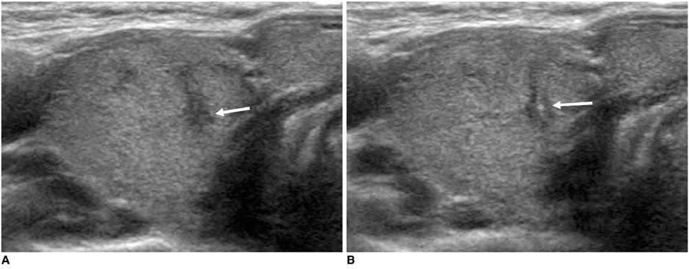
Fig. 1 Imaging findings are shown for solitary papillary thyroid microcarcinoma (approximately 3.0 mm in maximum diameter). A. Transverse sonogram of right lobe of thyroid shows presence of markedly hypoechoic nodule (arrow) with ill-defined margin and taller-than-wide shape. B. Transverse sonogram shows echogenic dot (arrow) within same nodule due to needle tip during US-fine needle aspiration biopsy.
Reference
-
1. Hedinger C, Williams ED, Sobin LH. World Health Organization. Histological typing of thyroid tumours. International histological classification of tumours. 1988. 2nd ed. Berlin: Springer-Verlag.2. Leenhardt L, Hejblum G, Farnc B, Fediaevsky LD, Delbot T, Le Guillouzic D, et al. Indications and limits of ultrasound-guided cytology in the management of nonpalpable thyroid nodules. J Clin Endocrinol Metab. 1999. 84:24–28.3. Kim SJ, Kim EK, Park CS, Chung WY, Oh KK, Yoo HS. Ultrasound-guided fine-needle aspiration biopsy in nonpalpable thyroid nodules: is it useful in infracentimetric nodules? Yonsei Med J. 2003. 44:635–640.4. Nam-Goong IS, Kim HY, Gong G, Lee HK, Hong SJ, Kim WB, et al. Ultrasonography-guided fine-needle aspiration of thyroid incidentaloma: correlation with pathological findings. Clin Endocrinol (Oxf). 2004. 60:21–28.5. Baloch ZW, Sack MJ, Yu GH, Livolsi VA, Gupta PK. Fine-needle aspiration of thyroid: an institutional experience. Thyroid. 1998. 8:565–569.6. Gharib H, Papini E, Valcavi R, Baskin HJ, Crescenzi A, Dottorini ME, et al. American Association of Clinical Endocrinologists and Associazione Medici Endorinologi medical guidelines for clinical practice for the diagnosis and management of thyroid nodules. Endocr Pract. 2006. 12:63–102.7. Cooper DS, Doherty GM, Haugen BR, Kloos RT, Lee SL, Mazzaferri EL, et al. Management guidelines for patients with thyroid nodules and differentiated thyroid cancer. Thyroid. 2006. 16:109–142.8. Frates MC, Benson CB, Charboneau JW, Cibas ES, Clark OH, Coleman BG, et al. Management of thyroid nodules detected at US: Society of Radiologists in Ultrasound consensus conference statement. Radiology. 2005. 237:794–800.9. Burman KD. Micropapillary thyroid cancer: should we aspirate all nodules regardless of size? J Clin Endocrinol Metab. 2006. 91:2043–2046.10. Mazzaferri EL, Kloos RT. Clinical review 128: Current approaches to primary therapy for papillary and follicular thyroid cancer. J Clin Endocrinol Metab. 2001. 86:1447–1463.11. Baskin HJ, Duick DS. The endocrinologists' view of ultrasound guidelines for fine needle aspiration. Thyroid. 2006. 16:207–208.12. Roti E, Rossi R, Trasforini G, Bertelli F, Ambrosio MR, Busutti L, et al. Clinical and histological characteristics of papillary thyroid microcarcinoma: results of retrospective study in 243 patients. J Clin Endocrinol Metab. 2006. 91:2171–2178.13. Chow SM, Law SC, Chan JK, Au SK, Yau S, Lau WH. Papillary microcarcinoma of the thyroid-Prognostic significance of lymph node metastasis and multifocality. Cancer. 2003. 98:31–40.14. Ceresini G, Corcione L, Morganti S, Milli B, Bertone L, Prampolini R, et al. Ultrasound-guided fine-needle capillary biopsy of thyroid nodules, coupled with on-site cytologic review, improves results. Thyroid. 2004. 14:385–389.15. Ito Y, Uruno T, Nakano K, Takamura Y, Miya A, Kobayashi K, et al. An observation trial without surgical treatment in patients with papillary microcarcinoma of the thyroid. Thyroid. 2003. 13:381–387.
- Full Text Links
-
- Actions
-
Cited
- CITED
-
- Close
- Share
-
- Similar articles
-
- Ultrasound-Guided Fine-Needle Aspiration Biopsy in Infracentimetric Thyroid Nodules
- Ultrasonography-guided Fine-needle Aspiration for Solid Thyroid Nodules Less than 5 mm in the Largest Diameter: Comparison in Diagnostic Adequacy and Accuracy According to Nodule Size
- Diagnosis of Parathyroid Adenoma Detected during Thyroid Ultrasound: The Role of Parathormone Measurement in Fine-Needle Aspiration Washout
- An Analysis of the Ultrasound Findings of False Negative Cases for an Initial Ultrasound-guided Fine Needle Aspiration Biopsy (FNAB)
- Ultrasound-guided Fine-needle Aspiration Biopsy of Thyroid Nodules: Comparison of the Pain Scale according to the Application of Local Anesthesia
