Adventitial Cystic Disease of the Femoral Vein: a Case Report with the CT Venography
- Affiliations
-
- 1Department of Radiology, University of Konyang College of Medicine, Daejeon, Korea.
- 2Department of Radiology, Gachon University Gil Hospital, Incheon, Korea. bookdoo7@chollian.net
- 3Department of Radiology, Sam Hospital, Anyang, Korea.
- KMID: 1088684
- DOI: http://doi.org/10.3348/kjr.2009.10.1.89
Abstract
- Fewer than 20 cases of adventitial cystic disease of the vein have been reported in the worldwide literature. This small number of reported cases may be due not only to the disease's low incidence, but also to the difficulty in making the proper diagnosis. Many techniques have been used to investigate this disease, but venography has been the traditional diagnostic tool. In this report we present a case of adventitial cystic disease that was well demonstrated by CT venography.
MeSH Terms
Figure
-
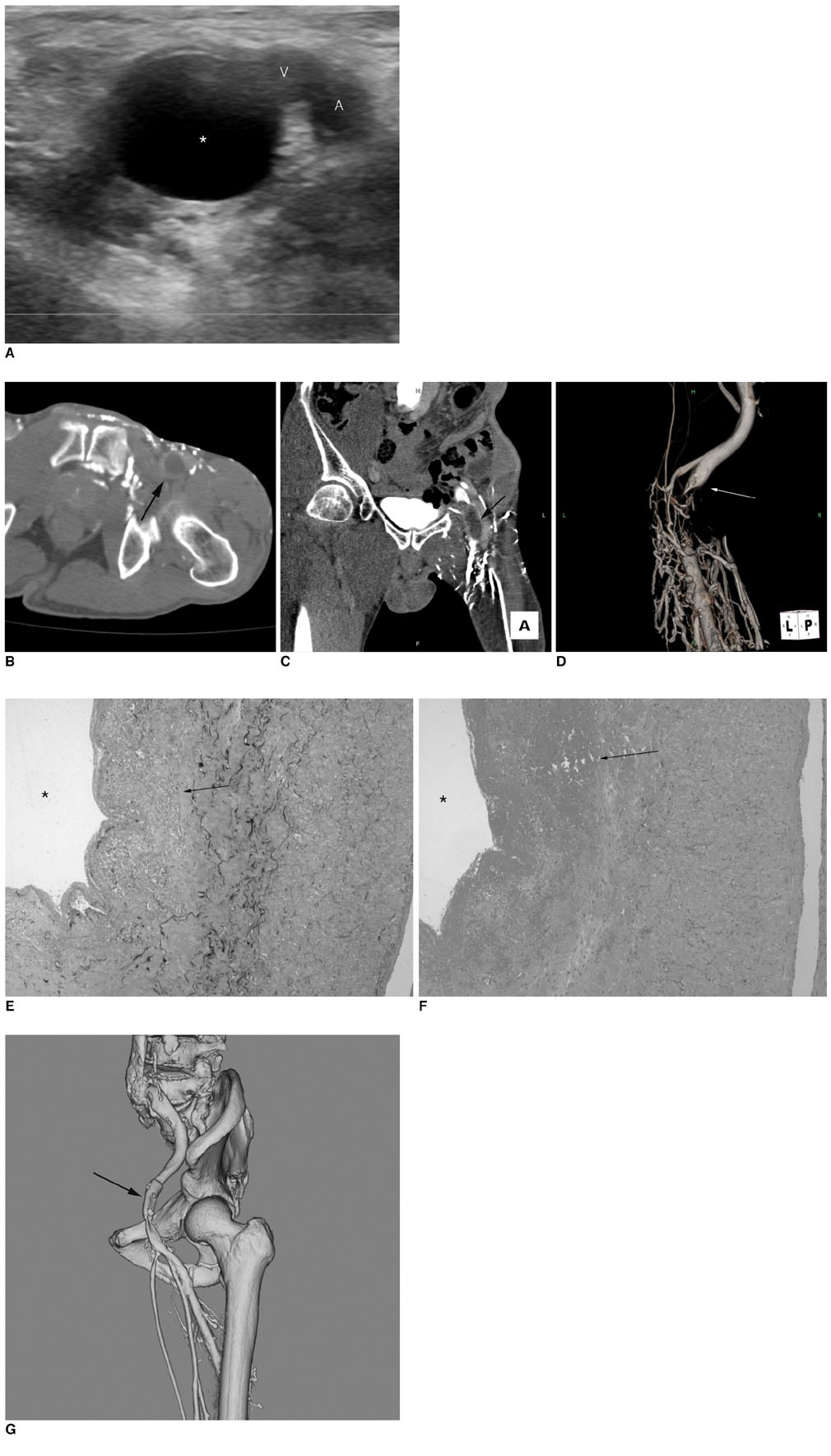
Fig. 1 US of adventitial cystic disease in 69-year-old man. A. US showed relationship between cystic mass and left common femoral vein. Cystic mass (*) that contained hypoechoic materials is attached to medial side of left common femoral vein (v). B. Venous phase axial CT scan shows approximately 1.7 cm sized round, nonenhancing cystic mass (long arrow) compressing left common femoral vein with dilated medial circumflex femoral and obturator veins, which provide collateral circulation. C. Coronal reconstruction image shows low attenuation nonenhancing mass (white arrow) in left common femoral vein. D. Posterior view of three dimensional volume rendering image of left common femoral vein showing near complete obstruction of left common femoral vein (white arrow) with formation of multiple collateral vessels. E. Photomicrograph of excised left common femoral vein. ×40; Elastin Van Gieson stain reveals widening of adventitial layer of vein and replacement of elastic tissue by fibrous connective tissue (arrow), which is all consistent with cystic adventitial disease. Cyst was removed (asterisk). F. Photomicrograph of excised left common femoral vein. ×40; Hematoxylin & Eosin stain shows adventitial hemorrhage and fibrin deposition within widened adventitial layer of vein (arrow). * = cyst G. Postoperative CT venography of patient's adventitial cystic disease. Lateral view of three dimensional volume rendering image showing that left common femoral vein (arrow) runs through previous cystic mass site and multiple collateral vessels have disappeared.
Reference
-
1. Wilbur AC, Woelfel GF, Meyer JP, Flanigan DP, Spigos DG. Adventitial cystic disease of the popliteal artery. Radiology. 1985. 155:63–64.2. Atkins HJB, Key JA. A case of myxomatous tumour arising in the adventitia of the left external iliac artery. Br J Surg. 1947. 34:426–427.3. Velasquez G, Zollikofer C, Nath HP, Barreto A, Castaneda-Zuniga W, Formanek A, et al. Cystic arterial adventitial degeneration. Radiology. 1980. 134:19–21.4. Do DD, Braunschweig M, Baumgartner I, Furrer M, Mahler F. Adventitial cystic disease of the popliteal artery: percutaneous US-guided aspiration. Radiology. 1997. 203:743–746.5. Dix FP, McDonald M, Obomighie J, Chalmers N, Thompson D, Benbow EW, et al. Cystic adventitial disease of the femoral vein presenting as deep vein thrombosis: a case report and review of the literature. J Vasc Surg. 2006. 44:871–874.6. Maldonado-Fernandez N, Lopez-Espada C, Moreno-Escobar J, Martinez-Gamez J, Rodriguez-Morata A, Garcia-Rospide V. Recurring adventitial cyst in the left external iliac vein. EJVES Extra. 2004. 8:10–14.7. Cho K, Shin TB. A case of adventitial cystic disease of the femoral vein. J Korean Soc Vasc Surg. 2005. 21:186–189. [Korean].8. Gagnon J, Doyle DL. Adventitial cystic disease of common femoral artery. Ann Vasc Surg. 2007. 21:84–86.9. Fukui S, Paraskevas N, Lafaurie C, Soury P, Gigou F, Petit MD, et al. Cystic formation compressing the femoral vein: synovial hip joint or adventitial cyst. EJVES Extra. 2004. 8:1–4.10. Jasinski RW, Masselink BA, Partridge RW, Deckinga BG, Bradford PF. Adventital cystic disease of the popliteal artery. Radiology. 1987. 163:153–155.
- Full Text Links
-
- Actions
-
Cited
- CITED
-
- Close
- Share
-
- Similar articles
-
- Adventitial Cystic Disease of the Common Femoral Vein Mimicking Deep Venous Thrombosis: A Case Report
- Adventitial Cystic Disease of the Superficial Femoral Vein without a Joint Connection: A Case Report
- Adventitial Cystic Disease of the Common Femoral Artery: A Case Report and Literature Review
- A Case of Adventitial Cystic Disease of the Femoral Vein
- Adventitial Cystic Disease of the Left External Iliac Vein: A Case Report
