The Clinical Usefulness of 18F-FDG PET/CT for the Evaluation of Lymph Node Metastasis in Periorbital Malignancies
- Affiliations
-
- 1Department of Otorhinolaryngology-Head and Neck Surgery, Samsung Medical Center, Sungkyunkwan University School of Medicine, Seoul, Korea. hansin.jeong@samsung.com
- 2Department of Ophthalmology, Samsung Medical Center, Sungkyunkwan University School of Medicine, Seoul, Korea.
- 3Department of Nuclear Medicine, Samsung Medical Center, Sungkyunkwan University School of Medicine, Seoul, Korea.
- 4Department of Radiology, Samsung Medical Center, Sungkyunkwan University School of Medicine, Seoul, Korea.
- 5Department of Pathology, Samsung Medical Center, Sungkyunkwan University School of Medicine, Seoul, Korea.
- KMID: 1088671
- DOI: http://doi.org/10.3348/kjr.2009.10.1.1
Abstract
OBJECTIVE
The aim of this study was to assess the clinical role of 18F-FDG PET/CT for the evaluation of lymph node metastasis in periorbital malignancies, compared with CT alone.
MATERIALS AND METHODS
We analyzed eighteen PET/CT and CT scans in 15 patients with biopsy-proven periorbital malignancies. We compared the diagnostic capabilities of PET/CT and CT with regard to nodal metastasis by level-by-level analysis and by N staging prediction. The reference standards were surgical pathology (n = 7) from dissected lymph node specimens and the results from radiological follow-up (n = 11, mean 20.5 months; range 10-52 months). Moreover, any changes in patient care as prompted by PET/CT were recorded and compared with treatment planning for CT alone.
RESULTS
PET/CT had a sensitivity of 100%, while CT had a sensitivity of 57% (p = 0.03) for nodal metastasis by level-by-level analysis. PET/CT had a specificity of 97%, positive predictive value of 93%, negative predictive value of 100%, and diagnostic accuracy of 98%, while the CT values for these same parameters were 97%, 89%, 82%, and 84%, respectively. PET/CT correctly predicted N staging with an accuracy of 100%, while CT was only 83% accurate (p = 0.01). Regarding the impact on patient care, the extent of surgery for regional lymph nodes and the treatment decision were modified by PET/CT in 39% of patients.
CONCLUSION
PET/CT could provide useful information in the management of regional lymph node metastases in patients with periorbital malignancies.
MeSH Terms
-
Adult
Aged
Contrast Media
Eye Neoplasms/pathology/*secondary
Eyelid Neoplasms/pathology/secondary
Female
Fluorodeoxyglucose F18/*diagnostic use
Humans
Iohexol/analogs & derivatives/diagnostic use
Lacrimal Apparatus Diseases/pathology
Lymphatic Metastasis/diagnosis
Male
Middle Aged
*Positron-Emission Tomography
Radiopharmaceuticals/*diagnostic use
Sensitivity and Specificity
*Tomography, X-Ray Computed/methods
Figure
-

Fig. 1 65-year-old male patient with sebaceous carcinoma in upper eyelid. A. On contrast-enhanced CT, no definite lesion is detected around parotid gland. B, C. On PET/CT, malignant lesion with high glucose uptake of SUV 4.7 is diagnosed at same site (arrows). According to fused image, malignant lesion is located around parotid gland.
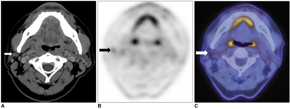
Fig. 2 56-year-old male patient with sebaceous carcinoma in upper eyelid. A. On contrast-enhanced CT, upper jugular lymph node behind angle of mandible is considered benign, with pattern of low enhancement and oval shape (arrow). B, C. On PET/CT, same lesion showed asymmetrical glucose uptake with SUV 2.2 and is diagnosed as metastatic lymph node (arrows). As result, surgical fields were extended to include specific lymph node, which was pathologically proven to have metastatic lesion.
Reference
-
1. Limawararut V, Leibovitch I, Sullivan T, Selva D. Periocular squamous cell carcinoma. Clin Experiment Ophthalmol. 2007. 35:174–185.2. Cook BE Jr, Bartley GB. Treatment options and future prospects for the management of eyelid malignancies: an evidence-based update. Ophthalmology. 2001. 108:2088–2098.3. Donaldson MJ, Sullivan TJ, Whitehead KJ, Williamson RM. Squamous cell carcinoma of the eyelids. Br J Ophthalmol. 2002. 86:1161–1165.4. Reifler DM, Hornblass A. Squamous cell carcinoma of the eyelid. Surv Ophthalmol. 1986. 30:349–365.5. Kass LG, Hornblass A. Sebaceous carcinoma of the ocular adnexa. Surv Ophthalmol. 1989. 33:477–490.6. Jeong HS, Son YI, Baek CH. The pattern of lymphatic metastasis of malignant tumors in the periorbital area. Am J Otolaryngol. 2006. 27:5–8.7. Nowak B, Di Martino E, Jänicke S, Cremerius U, Adam G, Zimny M, et al. Diagnostic evaluation of malignant head and neck cancer by F-18-FDG PET compared to CT/MRI. Nuklearmedizin. 1999. 38:312–318.8. Branstetter BF 4th, Blodgett TM, Zimmer LA, Snyderman CH, Johnson JT, Raman S, et al. Head and neck malignancy: is PET/CT more accurate than PET or CT alone? Radiology. 2005. 235:580–586.9. Schoder H, Yeung HW, Gonen M, Kraus D, Larson SM. Head and neck cancer: clinical usefulness and accuracy of PET/CT image fusion. Radiology. 2004. 231:65–72.10. Zimmer LA, Branstetter BF, Nayak JV, Johnson JT. Current use of 18F-fluorodeoxyglucose positron emission tomography and combined positron emission tomography and computed tomography in squamous cell carcinoma of the head and neck. Laryngoscope. 2005. 115:2029–2034.11. Jeong HS, Chung MK, Baek CH, Choi JY, Son YI, Kim HJ, et al. Combined 18F-FDG PET/CT imaging for the initial evaluation of glottic cancer. Clin Exp Otorhinolaryngol. 2008. 1:35–40.12. Donaldson MJ, Pulido JS, Mullan BP, Inwards DJ, Cantrill H, Johnson MR, et al. Combined positron emission tomography/computed tomography for evaluation of presumed choroidal metastases. Clin Experiment Ophthalmol. 2006. 34:846–851.13. Lane KA, Bilyk JR. Preliminary study of positron emission tomography in the detection and management of orbital malignancy. Ophthal Plast Reconstr Surg. 2006. 22:361–365.14. Roe RH, Finger PT, Kurli M, Tena LB, Iacob CE. Whole-body positron emission tomography/computed tomography imaging and staging of orbital lymphoma. Ophthalmology. 2006. 113:1854–1858.15. Valenzuela AA, Allen C, Grimes D, Wong D, Sullivan TJ. Positron emission tomography in the detection and staging of ocular adnexal lymphoproliferative disease. Ophthalmology. 2006. 113:2331–2337.16. Wild D, Eyrich GK, Ciernik IF, Stoeckli SJ, Schuknecht B, Goerres GW. In-line (18)F-fluorodeoxyglucose positron emission tomography with computed tomography (PET/CT) in patients with carcinoma of the sinus/nasal area and orbit. J Craniomaxillofac Surg. 2006. 34:9–16.17. Kim KH, Sung MW, Yun JB, Han MH, Baek CH, Chu KC, et al. The significance of CT scan or MRI in the evaluation of salivary gland tumors. Auris Nasus Larynx. 1998. 25:397–402.18. Jeong HS, Chung MK, Son YI, Choi JY, Kim HJ, Ko YH, et al. Role of 18F-FDG PET/CT in management of high-grade salivary gland malignancies. J Nucl Med. 2007. 48:1237–1244.19. Roh JL, Ryu CH, Choi SH, Kim JS, Lee JH, Cho KJ, et al. Clinical utility of 18F-FDG PET for patients with salivary gland malignancies. J Nucl Med. 2007. 48:240–246.
- Full Text Links
-
- Actions
-
Cited
- CITED
-
- Close
- Share
-
- Similar articles
-
- Supraclavicular Lymph Node Metastasis from Various Malignancies: Assessment with 18F-Fluorodeoxyglucose Positron Emission Tomography/CT, Contrast-Enhanced CT and Ultrasound
- Reliability of 18F-Fluorodeoxyglucose Positron Emission Tomography/Computed Tomography in the Nodal Staging of Colorectal Cancer Patients
- Usefulness of 18F-FDG PET/CT in the evaluation of cervical lymph node metastasis in patients with oral cancer
- F18-fluorodeoxyglucose-positron emission tomography and computed tomography is not accurate in preoperative staging of gastric cancer
- Comparison of Neck CT and ¹â¸F-FDG PET-CT for Making the Preoperative Diagnosis of Lymph Node Metastasis in Papillary Thyroid Cancer
