Korean J Ophthalmol.
2008 Sep;22(3):155-158. 10.3341/kjo.2008.22.3.155.
The Relationship Between the Density of Lens and Liquefaction Time Using Liquefaction Device
- Affiliations
-
- 1Department of Ophthalmology and Visual Science, St. Mary's Hospital, College of Medicine, The Catholic University of Korea, Seoul, Korea. eyedoc@catholic.ac.kr
- KMID: 1070749
- DOI: http://doi.org/10.3341/kjo.2008.22.3.155
Abstract
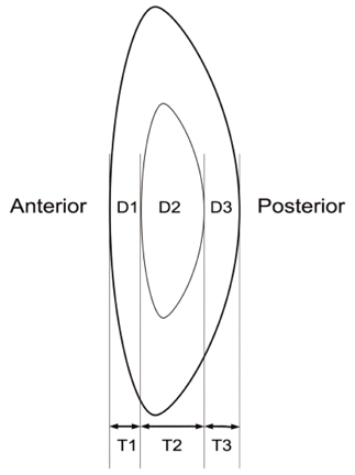
- PURPOSE: To investigate the effect of lens density on liquefaction time by using liquefaction device (AquaLase(R), Alcon Laboratories, TX, U.S.A.). METHODS: Cataract surgery using AquaLase(R) was performed on 47 eyes. With a Scheimpflug camera, the density and thickness of lens were measured in eye of each patient preoperatively. During surgery, liquefaction time and total number of pulses were recorded. The correlation of both density and thickness of lens with liquefaction time and total number of pulses was analyzed. RESULTS: The mean density of anterior cortex, nucleus, and posterior cortex was 112.45+/-42.1 computer compatible tapes (CCT), 76.5+/-22.7 CCT, and 70.9+/-52.2 CCT, respectively. The mean thickness was 0.97+/-0.30 mm, 2.76+/-0.54 mm, and 0.81+/-0.24 mm, respectively. The mean liquefaction time was 174.8+/-108.2 seconds. The mean total number of pulses was 4799+/-3007.There was no significant difference between the density of each area of lens (anterior cortex, nucleus, posterior cortex, and total lens) and liquefaction time (p>0.05), and between the thickness of each area of lens and liquefaction time (p>0.05). There was no significant difference between the density of each area of lens and total number of pulses (p>0.05), and between the thickness of each area of lens and total number of pulses (p>0.05). CONCLUSIONS: When extraction of soft to moderate density cataract was performed with AquaLase(R), liquefaction time and total number of pulse did not correlate to the density and thickness of lens.
MeSH Terms
Figure
Reference
-
1. Dick HB, Kohnen T, Jacobi FK, Jacobi KW. Long-term endothelial cell loss following phacoemulsification through a temporal clear corneal incision. J Cataract Refract Surg. 1996. 22:63–71.2. Hayashi K, Hayashi H, Nakao F, Hayashi F. Risk factors for corneal endothelial injury during phacoemulsification. J Cataract Refract Surg. 1996. 20:1079–1084.3. Qian W, Sodenberg P, Chen E, Philipson B. Universal opacity standard for Scheimpflug photography. Ophthalmic Res. 2000. 32:292–298.4. Mackool RJ, Brint SF. AquaLase : a new technology for cataract extraction. Curr Opin Ophthalmol. 2004. 15:40–43.5. Ogino K, Koda F, Miyaki K. Damage to cultured corneal endothelium caused by ultrasound during phacoemulsification. Nippon Ganka Gakkai Zasshi. 1993. 97:1286–1291.
- Full Text Links
- Actions
-
Cited
- CITED
-
- Close
- Share
- Similar articles
-
- Studies on Liquefaction of Human Semen
- The Relationship with the Density of Lens Nucleus and Phacoemulsification Time
- The Relationship of the Lens Density with the Lens Thickness and the Anterior Chamber Depth
- Splenic Liquefaction after Splenic Artery Embolization
- Semen Analysis in Husbands of Infertile Couples


