J Vet Sci.
2011 Mar;12(1):91-94. 10.4142/jvs.2011.12.1.91.
Computed tomographic evaluation of abdominal fat in minipigs
- Affiliations
-
- 1Department of Veterinary Medical Imaging, College of Veterinary Medicine and Research Institute for Veterinary Science, Seoul National University, Seoul 151-742, Korea. mcchoi@snu.ac.kr
- 2College of Veterinary Medicine, Chungbuk National University, Cheongju 361-763, Korea.
- KMID: 1067342
- DOI: http://doi.org/10.4142/jvs.2011.12.1.91
Abstract
- Computed tomography (CT) exams were conducted to determine the distribution of abdominal fat identified based on the CT number measured in Hounsfield Units (HU) and to measure the volume of the abdominal visceral and subcutaneous fat in minipigs. The relationship between the CT-based fat volumes of several vertebral levels and the entire abdomen and anthropometric data including the sagittal abdominal diameter and waist circumference were evaluated. Moreover, the total fat volumes at the T11, T13, L3, and L5 levels were compared with the total fat volume of the entire abdomen to define the landmark of abdominal fat distribution. Using a single-detector CT, six 6-month-old male minipigs were scanned under general anesthesia. Three radiologists then assessed the HU value of visceral and subcutaneous abdominal fat by drawing the region of interest manually at the T11, T13, L1, L3, and L5 levels. The CT number and abdominal fat determined in this way by the three radiologists was found to be correlated (intra-class coefficient = 0.9). The overall HU ranges for the visceral and subcutaneous fat depots were -147.47 to -83.46 and -131.62 to -90.97, respectively. The total fat volume of the entire abdomen was highly correlated with the volume of abdominal fat at the T13 level (r = 0.97, p < 0.0001). These findings demonstrate that the volume of abdominal adipose tissue measured at the T13 level using CT is a strong and reliable predictor of total abdominal adipose volume.
MeSH Terms
Figure
-
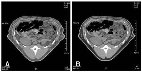
Fig. 1 Measurement of total (A, area of vertical stripes) and visceral (B) abdominal adipose tissue on the cross-sectional image at L3 level by thresholding using a computed tomography range of -147.47 to -83.46. To measure the volume of visceral adipose tissue, the region of interest is drawn manually surrounding the visceral cavity (B, shaded area within white line). The volume of subcutaneous adipose tissue is determined by subtracting the volume of visceral adipose tissue from that of the total adipose tissue.

Fig. 2 Mean Hounsfield unit (HU) values of abdominal fat at different levels (mean ± SD). The HU value of visceral fat was significantly lower than that of the subcutaneous fat at the L1, L3 and L5 levels.
Reference
-
1. Deschênes D, Couture P, Dupont P, Tchernof A. Subdivision of the subcutaneous adipose tissue compartment and lipid-lipoprotein levels in women. Obes Res. 2003. 11:469–476.
Article2. Fujioka S, Matsuzawa Y, Tokunaga K, Tarui S. Contribution of intra-abdominal fat accumulation to the impairment of glucose and lipid metabolism in human obesity. Metabolism. 1987. 36:54–59.
Article3. German AJ, Holden SL, Moxham GL, Holmes KL, Hackett RM, Rawlings JM. A simple, reliable tool for owners to assess the body condition of their dog or cat. J Nutr. 2006. 136:2031S–2033S.
Article4. Greenfield JR, Samaras K, Chisholm DJ, Campbell LV. Regional intra-subject variability in abdominal adiposity limits usefulness of computed tomography. Obes Res. 2002. 10:260–265.
Article5. Ishioka K, Okumura M, Sagawa M, Nakadomo F, Kimura K, Saito M. Computed tomographic assessment of body fat in beagles. Vet Radiol Ultrasound. 2005. 46:49–53.
Article6. Laflamme D. Development and validation of a body condition score system for cats: A clinical tool. Feline Pract. 1997. 25:13–18.7. Lambe NR, Conington J, McLean KA, Navajas EA, Fisher AV, Bünger L. In vivo prediction of internal fat weight in Scottish Blackface lambs, using computer tomography. J Anim Breed Genet. 2006. 123:105–113.
Article8. Lee K, Lee S, Kim YJ, Kim YJ. Waist circumference, dual-energy X-ray absortiometrically measured abdominal adiposity, and computed tomographically derived intra-abdominal fat area on detecting metabolic risk factors in obese women. Nutrition. 2008. 24:625–631.
Article9. Maurovich-Horvat P, Massaro J, Fox CS, Moselewski F, O'Donnell CJ, Hoffmann U. Comparison of anthropometric, area- and volume-based assessment of abdominal subcutaneous and visceral adipose tissue volumes using multi-detector computed tomography. Int J Obes (Lond). 2007. 31:500–506.
Article10. McEvoy FJ, Madsen MT, Strathe AB, Svalastoga E. Hounsfield Unit dynamics of adipose tissue and non-adipose soft tissues in growing pigs. Res Vet Sci. 2008. 84:300–304.
Article11. Park BJ, Kim YJ, Kim DH, Kim W, Jung YJ, Yoon JH, Kim CY, Cho YM, Kim SH, Lee KB, Jang JJ, Lee HS. Visceral adipose tissue area is an independent risk factor for hepatic steatosis. J Gastroenterol Hepatol. 2008. 23:900–907.
Article12. Rössner S, Bo WJ, Hiltbrandt E, Hinson W, Karstaedt N, Santago P, Sobol WT, Crouse JR. Adipose tissue determinations in cadavers-a comparison between cross-sectional planimetry and computed tomography. Int J Obes. 1990. 14:893–902.13. Tanaka K, Miyashiro I, Yano M, Kishi K, Motoori M, Seki Y, Noura S, Ohue M, Yamada T, Ohigashi H, Ishikawa O. Accumulation of excess visceral fat is a risk factor for pancreatic fistula formation after total gastrectomy. Ann Surg Oncol. 2009. 16:1520–1525.
Article14. Tokunaga K, Matsuzawa Y, Ishikawa K, Tarui S. A novel technique for the determination of body fat by computed tomography. Int J Obes. 1983. 7:437–445.15. Van der Kooy K, Seidell JC. Techniques for the measurement of visceral fat: a practical guide. Int J Obes Relat Metab Disord. 1993. 17:187–196.16. Wajchenberg BL. Subcutaneous and visceral adipose tissue: their relation to the metabolic syndrome. Endocr Rev. 2000. 21:697–738.
Article17. Yoshizumi T, Nakamura T, Yamane M, Waliul Islam AHM, Menju M, Yamasaki K, Arai T, Kotani K, Funahashi T, Yamashita S. Abdominal Fat: standardized technique for measurement at CT. Radiology. 1999. 211:283–286.
Article
- Full Text Links
-
- Actions
-
Cited
- CITED
-
- Close
- Share
-
- Similar articles
-
- Ultrasonography and CT Findings of Epigastric Hernia: 3 Case Report
- Computed Tomographic Findings and Clinical Observation of Spontaneous Intracerebral Hemorrhage
- Computed Tomographic Dacfyocystography using Rayvist(R)
- The correlation between simple anthropometric indices and abdominal visceral fat accumulation by computed tomography
- Usefulness of Computed Tomographic Angiography(CTA) in the Evaluation of Cerebral Aneurysms
