Korean J Radiol.
2004 Mar;5(1):72-74. 10.3348/kjr.2004.5.1.72.
Craniopharyngioma in the Temporal Lobe: A Case Report
- Affiliations
-
- 1Department of Radiology, Keimyung University, Daegu, Korea. chsohn@dsmc.or.kr
- 2Department of Pathology, Keimyung University, Daegu, Korea.
- 3Department of Neurosurgery, Keimyung University, Daegu, Korea.
- 4Department of Radiology, University of Calgary, Calgary, Alberta, Canada.
- KMID: 1066248
- DOI: http://doi.org/10.3348/kjr.2004.5.1.72
Abstract
- Herein, we report on an unusual case of craniopharyngioma arising in the temporal lobe with no prior history of surgery and with no connection to the craniopharyngeal duct. MR images showed a cystic tumor with a small solid portion. To the best of our knowledge, this is the first case of a craniopharyngioma occurring in the temporal lobe.
Keyword
MeSH Terms
Figure
-
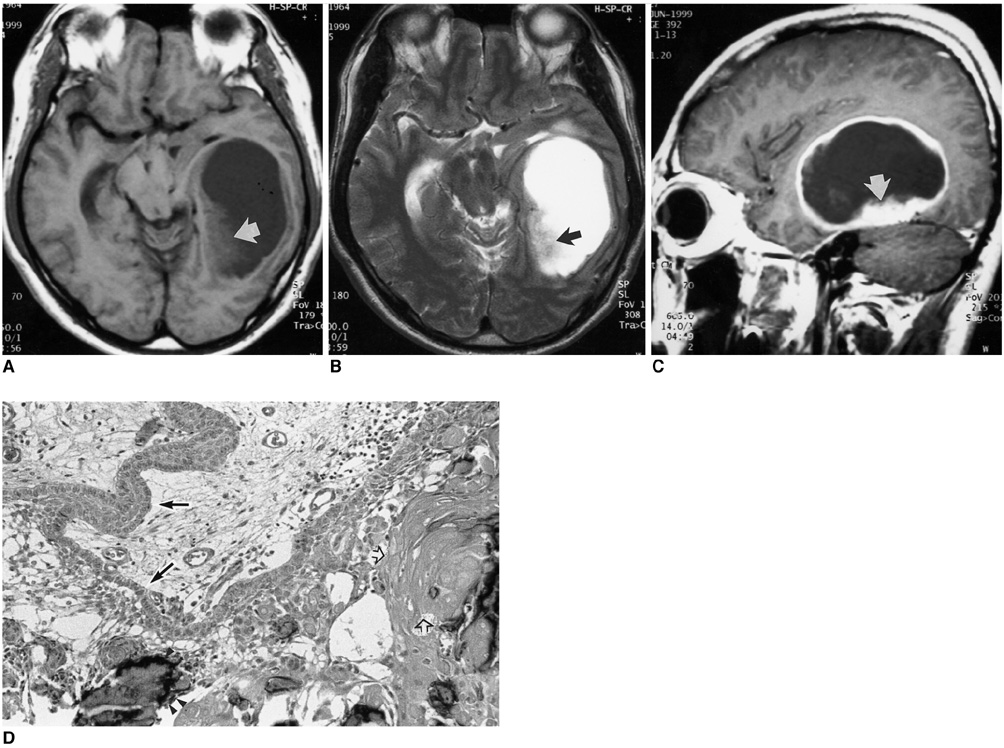
Fig. 1 Craniopharyngioma of the temporal lobe. Axial T1-weighted (A) and T2-weighted (B) MR images show a large cystic mass in the left temporal lobe, which contains small solid portions on its medial aspect (arrow). Sagittal contrast-enhanced T1-weighted (C) MR image shows thick peripheral rim enhancement, and well enhancing solid portion in the inferior part of the mass (arrow). Photomicrograph (D) shows classic adamantinomatous craniopharyngioma with peripheral palisades of epithelial cells (arrows), containing loose squamous cells separated by intercellular fluid. Delicate squamous epithelial cells contain wet keratin (open arrows) and calcification (arrowheads) (original magnification, ×100; hematoxylin-eosin staining).
Reference
-
1. Bock E. Beitrag zur pathologie der hypophyse. Virchow Arch Patholo Anat. 1924. 252:98–112.2. Benitez WI, Sartor KJ, Angtuaco EJC. Craniopharyngioma presenting as a nasopharyngeal mass: CT and MR findings. J Comput Assist Tomogr. 1988. 12:1068–1072.3. Linden CN, Martinez CR, Gonzalvo AA, Cahill DW. Intrinsic third ventricle craniopharyngioma: CT and MR findings. J Comput Assist Tomogr. 1989. 13:362–368.4. Lee JH, Kim CY, Kim DG, Jung HW. Postoperative ectopic seeding of craniopharyngioma: Case illustration. J Neurosurg. 1999. 90:796.5. Waga S, Morikawa A, Sakakura M. Craniopharyngioma with midbrain involvement. Arch Neurol. 1979. 36:319–320.6. Solarski A, Panke ES, Panke TW. Craniopharyngioma in the pineal gland. Arch Pathol Lab Med. 1978. 102:490–491.7. Sartoretti-Schefer S, Wichmann W, Aguzzi A, Valavanis A. MR differentiation of adamantinous and squamous-papillary craniopharyngiomas. AJNR Am J Neuroradiol. 1997. 18:77–87.8. Kanungo N, Just N, Black M, et al. Nasopharyngeal craniopharyngioma in an unusual location. AJNR Am J Neuroradiol. 1995. 16:1372–1374.9. Graziani N, Donnet A, Bugha TN, Dufour H, Figarella-Branger D, Grisoli F. Ectopic basisphenoidal craniopharyngioma: case report and review of the literature. Neurosurgery. 1994. 34:346–349.10. Gokalp HZ, Egemen N, Ildan F, Kadri B. Craniopharyngioma of the posterior fossa. Neurosurgery. 1991. 29:446–448.
- Full Text Links
-
- Actions
-
Cited
- CITED
-
- Close
- Share
-
- Similar articles
-
- Ictal Vomiting Associated with Temporal Lobe Epilepsy of Dominant Hemisphere
- Hemispheric laterality of temporal lobe epilepsy in relation to anxiety and depression
- Postictal MR Enhancement in a Patient with Lateral Temporal Lobe Epilepsy
- Temporal lobe epilepsy caused by intrahippocampal calcified cysticercus: a case report
- Three Patients with Nondominant Temporal Lobe Epilepsy Showing Ictal Spitting
