Comparison of Transvaginal Ultrasonography with Hysterosonography as a Screening Method in Patients with Abnormal Uterine Bleeding
- Affiliations
-
- 1Department of Radiology, Samsung Medical Center, Sungkyunkwan University School of Medicine. bhkim@smc.samsung.co.kr
- 2Department of Radiology, Asan Medical Center, University of Ulsan College of Medicine.
- KMID: 1066244
- DOI: http://doi.org/10.3348/kjr.2004.5.1.39
Abstract
OBJECTIVE
To assess the utility of hysterosonography (HS) as a screening method in patients with abnormal uterine bleeding. MATERIALS AND METHODS: We retrospectively reviewed transvaginal ultrasonography (TVS) and HS for 105 patients whose diagnosis was confirmed pathologically. All 105 patients were initially evaluated on the same day with both TVS and HS. On TVS and HS examination, endometrial cavitary lesions were classified as diffuse hyperplasia, endometrial polyp, endometrial cancer, uterine synechia and submucosal leiomyoma. Hysteroscopy with biopsy (n=35), curettage (n=60) or hysterectomy (n=10) was performed, and the results of TVS and HS examination were correlated with the pathological findings. RESULTS: The sensitivity and specificity were 79.0% and 45.8% for TVS, and 95.1% and 83.3% for HS, respectively. The positive and negative predictive values were 83.0% and 39.3% for TVS, and 95.1% and 83.3% for HS, respectively. Twenty-seven showed a discrepancy between the TVS and HS, and eight cases showed a discrepancy between HS and the pathologic diagnosis. CONCLUSION: TVS is a sensitive method to evaluate the endometrial cavitary lesions, but it often does not provide the physician with sufficient diagnostic information. With its higher sensitivities, specificities and positive and negative predictive values, HS can be better used than TVS in evaluating those patients with abnormal uterine bleeding.
Keyword
MeSH Terms
Figure
-
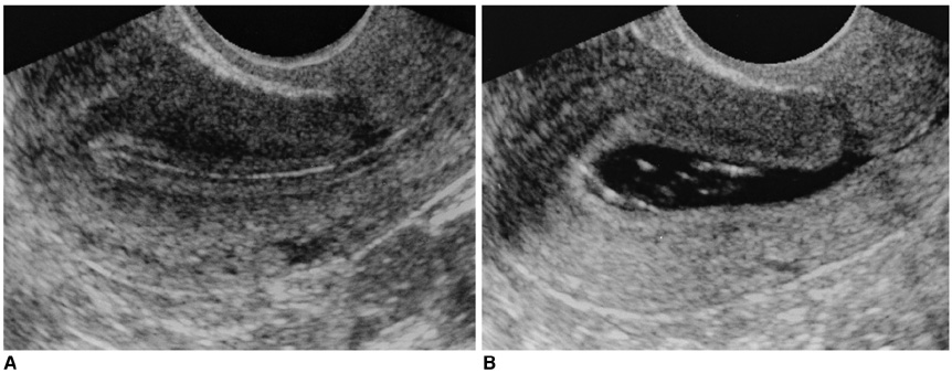
Fig. 1 A 42-year-old woman presented with dysmenorrhea and frequent vaginal spotting. A. Longitudinal image of transvaginal ultrasonography shows a thin smooth endometrium without any focal lesion. B. Longitudinal image of hysterosonography shows no abnormality of the endometrium. A small amount of debris in the endometrial cavity and a scar from previous Caesarean section in the anterior wall of the isthmic portion are seen.
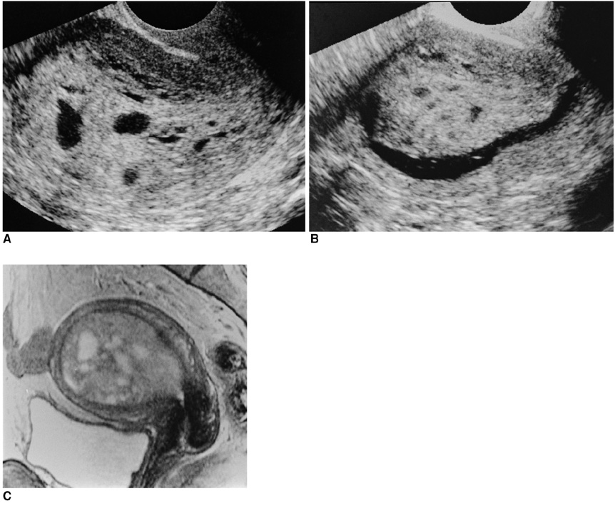
Fig. 2 A 45 year-old woman presented with abnormal uterine bleeding. A. Longitudinal image of transvaginal ultrasonography shows an extremely thickened endometrium and multiple cystic portions in the mass. B. Longitudinal image of hysterosonography shows a huge endometrial tumor, which was regarded as a polyp. C. T2-weighted sagittal image of the uterus shows about a 9 cm sized heterogeneous hyperintense mass confined in the endometrial cavity. Hysteroscopic removal was done and the diagnosis of endometrial polyp was confirmed in pathology.
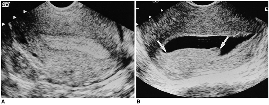
Fig. 3 A 48 year-old premenopausal woman presented with menorrhagia and dysfunctional uterine bleeding. A. Longitudinal image of transvaginal ultrasonography shows a homogeneously hyperechoic 12-mm-thick endometrium without any focal lesion, which looks normal. B. Longitudinal image of hysterosonography shows an elongated endometrial mass (arrows), which was regarded as endometrial hyperplasia. Curettage biopsy was done and an endometrial polyp was confirmed upon pathologic examination.
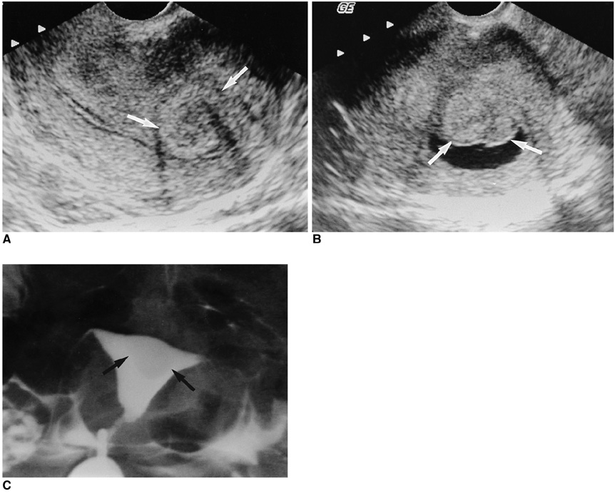
Fig. 4 A 37 year-old premenopausal woman presented with menorrhagia and dysfunctional uterine bleeding. A. Longitudinal image of transvaginal ultrasonography shows an intramural leiomyoma (arrows). B. Longitudinal image of hysterosonography shows a lobulated mass in a submucosal location (arrows). C. Upon hysterosalpingography, an intracavitary filling-defect (arrows) is seen. The diagnosis of leiomyoma was confirmed in pathology.
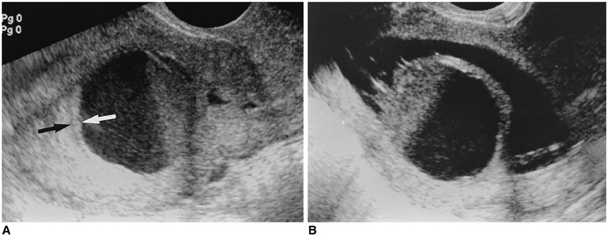
Fig. 5 A 48 year-old premenopausal woman presented with dysfunctional uterine bleeding. A. Longitudinal image of transvaginal ultrasonography shows hematometra with a fluid-debris level, and a cervical stenosis is suspected. A thin endometrium (arrows) is seen, which looks normal. B. Longitudinal image of hysterosonography shows a large endometrial mass, which is near totally necrotic. Total hemorrhagic degeneration of a submucosal myoma was suspected. Hysterectomy was done and adenomyoma was confirmed with pathology testing.
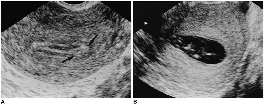
Fig. 6 A 58 year-old postmenopausal woman presented with vaginal spotting. A. Longitudinal image of transvaginal ultrasonography shows a small hyperechoic nodule (arrows) in the endometrium. It was regarded as a small polyp. B. Longitudinal image of hysterosonography shows a thin endometrium without any focal lesion. On the 1 month follow-up transvaginal ultrasonography, the small nodule disappeared and the vaginal spotting disappeared as well.
Reference
-
1. Bree RL, Bowerman RA, Bohm-Velez M, et al. US evaluation of the uterus in patients with postmenopausal bleeding: A positive effect on diagnostic decision making. Radiology. 2000. 216:260–264.2. Dubinsky TJ, Parvey HR, Maklad N. The role of transvaginal sonography and endometrial biopsy in the evaluation of peri- and postmenopausal bleeding. AJR Am J Roentgenol. 1997. 169:145–149.3. Fleischer AC. Sonographic assessment of endometrial disorders. Semin Ultrasound CT MR. 1999. 20:259–266.4. Lee EJ, Lee HM, Kwon HC, Joo HJ. Usefulness of sonohysterography in the differentiation of endometrial and endometrial cavity abnormalities: comparison with transvaginal sonography. J Korean Soc Med Ultrasound. 1995. 14:175–181.5. Lee EJ, Kim JM, Ryu HS. Usefulness of sonohysterography in differentiating endometrial cancer from endometrial hyperplasia. J Korean Soc Med Ultrasound. 1999. 18:91–97.6. Saidi MH, Sadler RK, Theis VD, Akright BD, Farhart SA, Villanueva GR. Comparison of sonography, sonohysterography, and hysteroscopy for evaluation of abnormal uterine bleeding. J Ultrasound Med. 1997. 16:587–591.7. O'Connell LP, Fries MH, Zeringue E, Brehm W. Triage of abnormal postmenopausal bleeding: a comparison of endometrial biopsy and transvaginal sonohysterography versus fractional curettage with hysteroscopy. Am J Obstet Gynecol. 1998. 178:956–961.8. Laifer-Narin SL, Ragavendra N, Lu DS, Sayre J, Perrella RR, Grant EG. Transvaginal saline hysterosonography: characteristics distinguishing malignant and various benign conditions. AJR Am J Roentgenol. 1999. 172:1513–1520.9. Bree RL. Ultrasound of the endometrium: facts, controversies, and future trends. Abdom Imaging. 1997. 22:557–568.10. Timmerman D, Deprest J, Bourne T, Van den Berghe I, Collins WP, Vergote I. A randomized trial on the use of ultrasonography or office hysteroscopy for endometrial assessment in postmenopausal patients with breast cancer who were treated with tamoxifen. Am J Obstet Gynecol. 1998. 179:62–70.11. Becker E Jr, Lev-Toaff AS, Kaufman EP, Halpern EJ, Edelweiss MI, Kurtz AB. The added value of transvaginal sonohysterography over transvaginal sonography alone in women with known or suspected leiomyoma. J Ultrasound Med. 2002. 21:237–247.12. Cullinan JA, Fleischer AC, Kepple DM, Arnold AL. Sonohysterography: a technique for endometrial evaluation. RadioGraphics. 1995. 15:501–514.13. Sohaey R, Woodward P. Sonohysterography: technique, endometrial findings, and clinical applications. Semin Ultrasound CT MR. 1999. 20:250–258.14. Laifer-Narin S, Ragavendra N, Parmenter EK, Grant EG. False-normal appearance of the endometrium on conventional transvaginal sonography: comparison with saline hysterosonography. AJR Am J Roentgenol. 2002. 178:129–133.
- Full Text Links
-
- Actions
-
Cited
- CITED
-
- Close
- Share
-
- Similar articles
-
- The efficacy of transvaginal sonography , saline infusion sonohysterography , and hysteroscopy for the evaluation of the patients with abnormal uterine bleeding
- The Usefulness of Vaginal Sonography in Evaluation of Endometrium in Postmenopausal Women
- Clinical Efficacy of Levonorgestrel-Releasing Intrauterine System (Mirena(R)) for Abnormal Uterine Bleeding
- Hysteroscopic Findings in Abnormal Uterine Bleeding
- Are Transvaginal Sonography and Color doppler findings Useful for the Diagnosis of Adenomyosis?
