Korean J Gastroenterol.
2011 Dec;58(6):346-349. 10.4166/kjg.2011.58.6.346.
Gastric Duplication Cyst Removed by Endoscopic Submucosal Dissection
- Affiliations
-
- 1Department of Internal Medicine, Pusan National University School of Medicine, Busan, Korea. doc0224@pusan.ac.kr
- 2Department of Pathology, Pusan National University School of Medicine, Busan, Korea.
- KMID: 1043334
- DOI: http://doi.org/10.4166/kjg.2011.58.6.346
Abstract
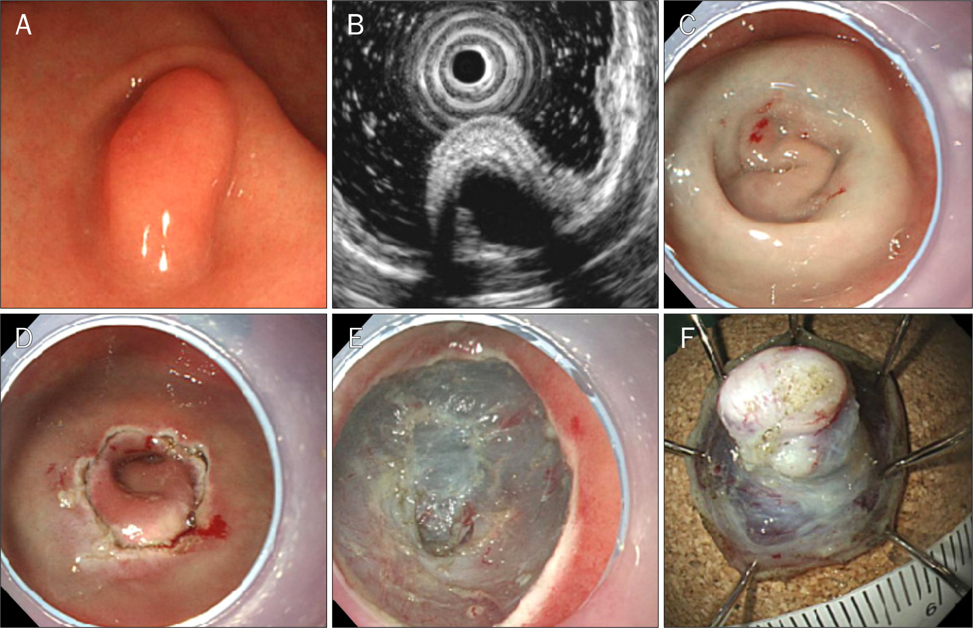
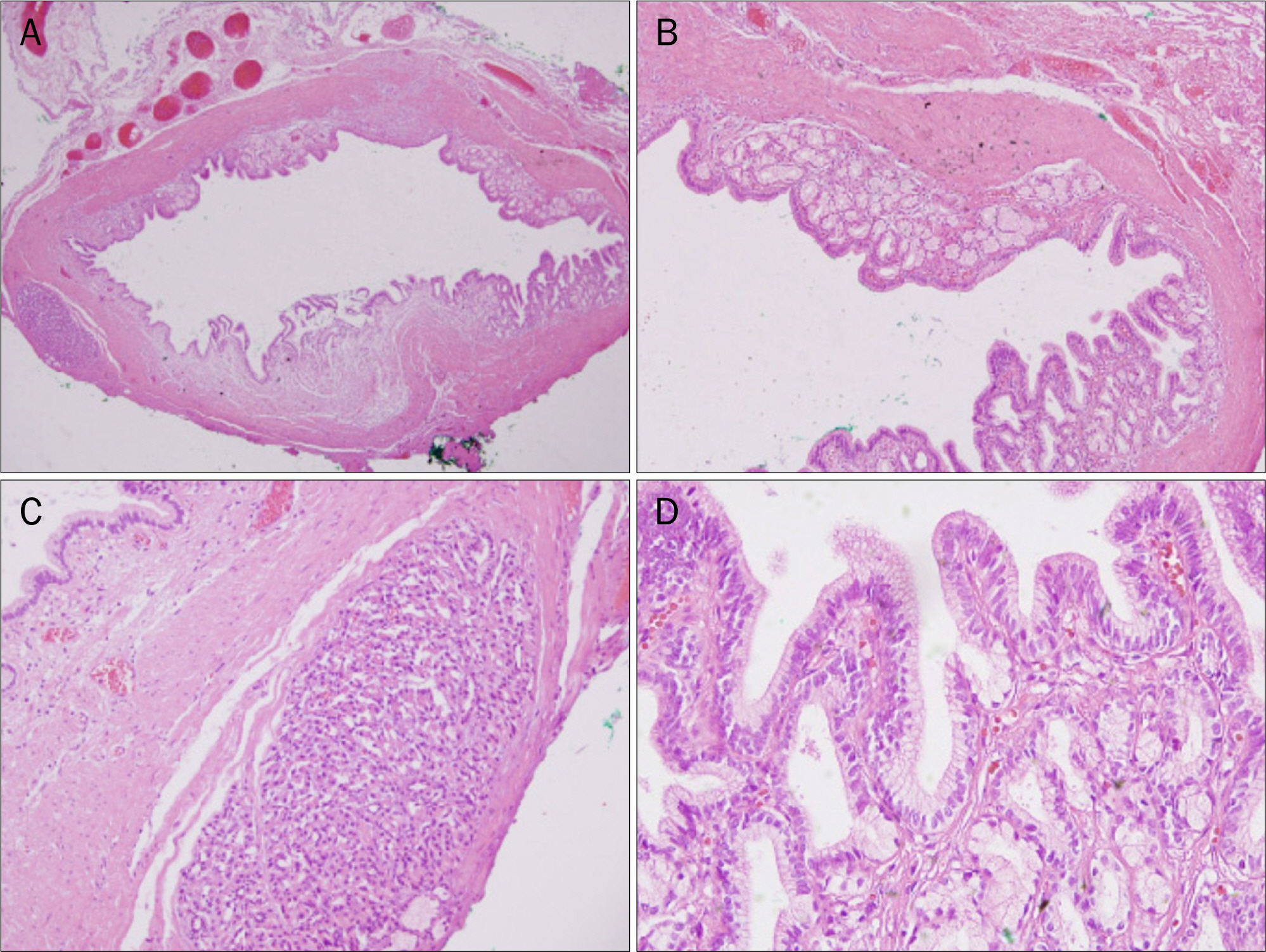
- Duplication cysts are uncommon congenital malformations that may occur anywhere throughout the alimentary tract. The stomach is an extremely rare site of occurrence. Here, we report a case of gastric duplication cyst initially presenting with a gastric submucosal tumor. A 28-year-old man complained of dyspepsia lasting 1 year and upper endoscopy revealed an ellipsoid submucosal tumor at the greater curvature of the antrum. We intended to use the injection-and-cut technique: however, after saline injection, the lesion was dented and impossible to grasp with a snare. Therefore, we decided to perform endoscopic submucosal dissection and removed the tumor without complication. Histopathology revealed a 0.6x0.6 cm-sized duplication cyst, and there has been no recurrence in 2 years.
Keyword
MeSH Terms
Figure
Reference
-
References
1. Kim DH, Kim JS, Nam ES, Shin HS. Foregut duplication cyst of the stomach. Pathol Int. 2000; 50:142–145.
Article2. Hsu HT, Hsing MT, Chen ML, Chen CJ. A gastric duplication cyst at the splenic hilum mimicking endometriosis clinically in a female adult. Chin Med J (Engl). 2009; 122:2079–2080.3. Chen PH, Lee JY, Yang SF, Wang JY, Lin JY, Chang YT. A retroperitoneal gastric duplication cyst mimicking a simple exophytic renal cyst in an adolescent. J Pediatr Surg. 2010; 45:e5–e8.
Article4. Lee YC, Kim YB, Kim JK, et al. Endoscopic treatment of a large gastric duplication cyst with hook-knife and snare (with video). Gastrointest Endosc. 2011; 73:1039–1040.
Article5. Murakami S, Isozaki H, Shou T, Sakai K, Toyota H. Foregut duplication cyst of the stomach with pseudostratified columnar cili-ated epithelium. Pathol Int. 2008; 58:187–190.
Article6. Kuraoka K, Nakayama H, Kagawa T, Ichikawa T, Yasui W. Adeno-carcinoma arising from a gastric duplication cyst with invasion to the stomach: a case report with literature review. J Clin Pathol. 2004; 57:428–431.
Article7. Bonacci JL, Schlatter MG. Gastric duplication cyst: a unique presentation. J Pediatr Surg. 2008; 43:1203–1205.
Article8. Macpherson RI. Gastrointestinal tract duplications: clinical, pathologic, etiologic, and radiologic considerations. Radiographics. 1993; 13:1063–1080.
Article9. Parra-Blanco A, Arnau MR, Nicolás-Pérez D, et al. Endoscopic submucosal dissection training with pig models in a Western country. World J Gastroenterol. 2010; 16:2895–2900.
Article
- Full Text Links
- Actions
-
Cited
- CITED
-
- Close
- Share
- Similar articles
-
- Endoscopic Submucosal Dissection in the Treatment of Patients With Papillary Early Gastric Cancer
- Gastric Inverted Hyperplastic Polyp Removed Using Endoscopic Submucosal Dissection
- Pathological Interpretation of Gastric Tumors in Endoscopic Submucosal Dissection
- “Intraluminal†Pyloric Duplication: A Case Report
- Adenocarcinoma Occurring in a Gastric Hyperplastic Polyp Treated with Endoscopic Submucosal Dissection


