Korean J Ophthalmol.
2010 Aug;24(4):237-239. 10.3341/kjo.2010.24.4.237.
Poliosis of Eyelashes as an Unusual Sign of a Halo Nevus
- Affiliations
-
- 1Department of Ophthalmology, Korea University Guro Hospital, Seoul, Korea. winona93@naver.com
- KMID: 949062
- DOI: http://doi.org/10.3341/kjo.2010.24.4.237
Abstract
- A 39-year-old man with poliosis of his lower eyelid lashes visited our clinic. He reported that his symptoms began with a few central lashes and then spread along the adjacent lashes during the ensuing 2 weeks. A pigmented nevus, approximately 4 mm in diameter, was identified just above the white lashes without surrounding skin depigmentation. No specific findings were identified with regard to the patient's general health or serologic and radiologic testing. Excisional biopsy of the pigmented nevus was performed. On histopathologic examination, infiltration of the dermis by numerous lymphocytes and melanophages was observed. The poliosis was ultimately diagnosed as a presenting sign of the halo phenomenon in the regressive stage of a melanocytic nevus.
Keyword
MeSH Terms
Figure
-
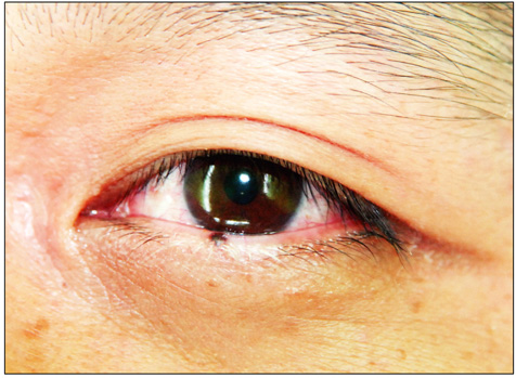
Fig. 1 Poliosis with a melanocytic nevus on the central eyelid.
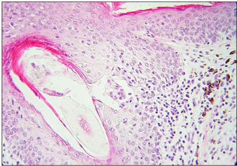
Fig. 2 No melanocytes are seen in the hair follicles, while melanocytes are observed in the overlying epidermis (H&E, ×40).

Fig. 3 Melanocytes (open arrow) in the dermis display maturation with progressive descent, much of the dermal component is obscured by a dense lymphohistiocytic infiltrate (arrow) (H&E, ×40).
Reference
-
1. Bertolino AP, Freedberg IM. Fitzpatrick TB, Eisen AZ, Wolff K, editors. Hair. Dermatology in general medicine. 2004. 4th ed. New York: McGraw-Hill;671–696.2. Moorthy RS, Inomata H, Rao NA. Vogt-Koyanagi-Harada syndrome. Surv Ophthalmol. 1995. 39:265–292.3. Goto H, Rao NA. Sympathetic ophthalmia and Vogt-Koyanagi-Harada syndrome. Int Ophthalmol Clin. 1990. 30:279–285.4. Wagoner MD, Albert DM, Lerner AB, et al. New observations on vitiligo and ocular disease. Am J Ophthalmol. 1983. 96:16–26.5. Chen CS, Wells J, Craig JE. Topical prostaglandin F(2 alpha) analog induced poliosis. Am J Ophthalmol. 2004. 137:965–966.6. Sotiriadis D, Lazaridou E, Patsatsi A, et al. Does halo nevus without halo exist? J Eur Acad Dermatol Venereol. 2006. 20:1394–1396.7. Barnhill RL, Fitzpatrick TB, Fandrey K, editors. Color atlas and synopsis of pigmented lesions. 1995. New York: McGraw-Hill;95–99.8. Mihara M, Nakayama H, Aki T, et al. Cutaneous nerves in cafe au lait spots with white halos in infants with neurofibromatosis: an electron microscopic study. Arch Dermatol. 1992. 128:957–961.9. Barnhill RL, Llewellyn K. Bolognia JL, Jorizzo JL, Rapini RP, editors. Benign melanocytic neoplasms. Dermatology. 2003. London: Mosby;1770.10. Elder DE, Elenitsas R, Murphy GF, et al. Elder DE, Elenitsas R, Johnson BL, Murphy GF, editors. Halo nevus. Lever's histopathology of the skin. 2005. 9th ed. Philadelphia: Lippincott Williams & Wilkins;747–749.11. Elder D, Elenitsas R. Elder D, Elenitsas R, Jaworsky C, Johnson B, editors. Benign pigmented lesions and malignant melanoma. Lever's histopathology of the skin. 1997. 8th ed. Philadelphia: Lippincott-Raven Publishers.12. Zack LD, Stegmeier O, Solomon LM. Pigmentary regression in a giant nevocellular nevus: a case report and a review of the subject. Pediatr Dermatol. 1988. 5:178–183.13. Kageshita T, Inoue Y, Ono T. Spontaneous regression of congenital melanocytic nevi without evidence of the halo phenomenon. Dermatology. 2003. 207:193–195.14. Guerra-Tapia A, Isarria MJ. Periocular vitiligo with onset around a congenital divided nevus of the eyelid. Pediatr Dermatol. 2005. 22:427–429.
