The "Mini-Perc" Technique of Percutaneous Nephrolithotomy with a 14-Fr Peel-away Sheath: 3-year Results in 72 Patients
- Affiliations
-
- 1Department of Radiology and Center for Imaging Science, Samsung Medical Center, Sungkyunkwan University School of Medicine, Seoul, Korea. swchoo@smc.samsung.co.kr
- 2Department of Urology, Samsung Medical Center, Sungkyunkwan University School of Medicine, Seoul, Korea.
- KMID: 753916
- DOI: http://doi.org/10.3348/kjr.2006.7.1.50
Abstract
OBJECTIVE
To assess the efficacy and safety of a "mini-perc" technique of percutaneous nephrolithotomy using a 14-Fr peel-away sheath for the removal of pyelocaliceal stones, and to determine appropriate inclusion criteria. MATERIALS AND METHODS: From July 1999 to June 2002, the medical records and radiographic images of 72 patients who underwent the "mini-perc" technique of percutaneous nephrolithotomy with a 14-Fr peel-away sheath, were reviewed to determine clinical history, stone characteristics, immediate stone free rate, final stone free rate after additional procedures, complications, and hospital stay. We also analyzed the effect of the longest stone diameter, the cumulative longest diameter of stones, the cumulative stone burden, and the stone density on the immediate stone free rate using a Fisher exact test. RESULTS: The only major complication, arterial bleeding, occurred in a patient with Child A liver cirrhosis and was successfully treated by embolization with coils and a gelatin sponge. The immediate stone free rate was 80.6 %, which was significantly influenced by stone diameter but not stone density. The mean hospital stay after the procedure was 3.97 days. CONCLUSION: The "mini-perc" technique of percutaneous nephrolithotomy, which uses the 14-Fr peel-away sheath, is a safe and effective modality for treating renal calculi.
MeSH Terms
Figure
-
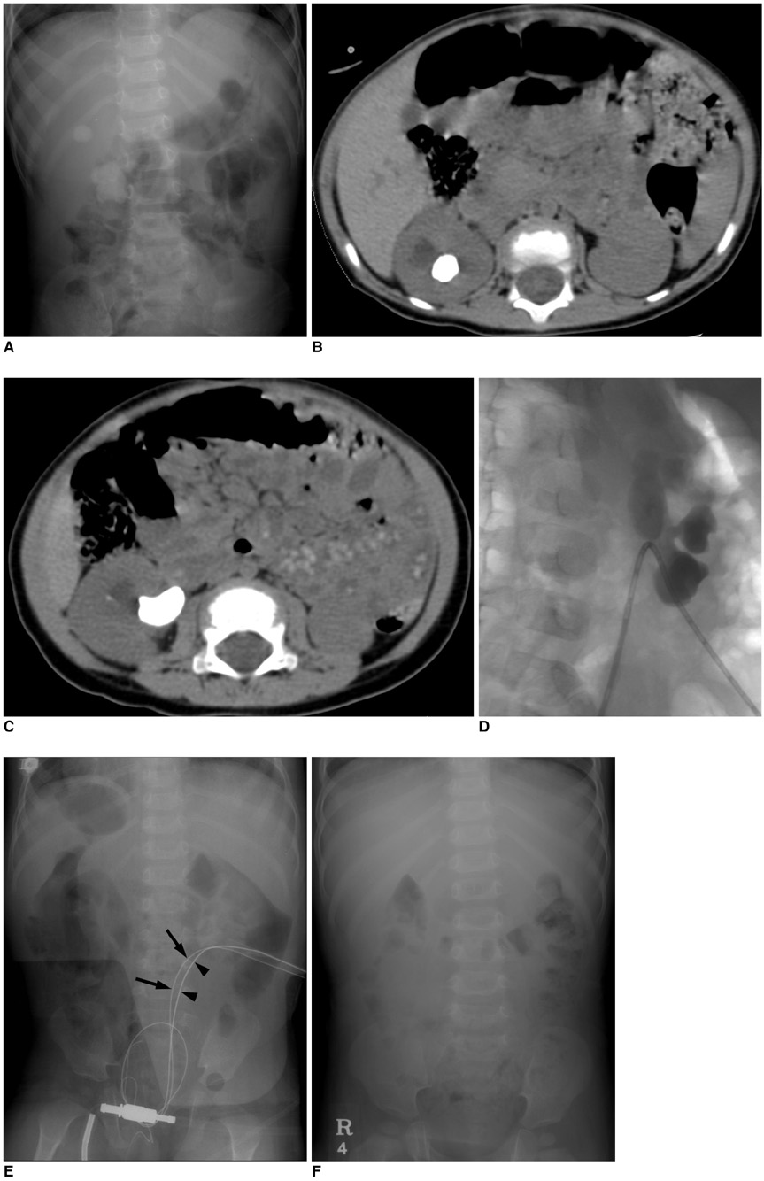
Fig. 1 A 17-month-old boy with upper pole calyceal and pelvic stones in the right kidney. A-C. Abdominal radiograph and non-enhanced helical CT scans (120 kVp, 160 mA, 2.5 mm collimation) show upper pole calyceal and pelvic stones in the right kidney. D. Nephrostomy was created toward the postero-inferior aspect of the renal stones. E. Intraoperative abdominal radiograph demonstrates a safety guide wire (arrows) and a working guide wire (arrowheads) inserted on prone position. F. One month after the procedure, renal stones are shown successfully removed on abdominal radiograph.
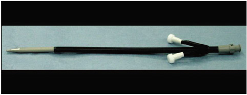
Fig. 2 A 14-Fr peel-away sheath for percutaneous access.
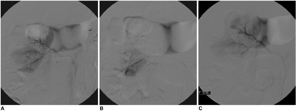
Fig. 3 A 65-year-old man with massive hematuria after the "mini-perc", who was diagnosed with Child A liver cirrhosis. A. Right renal arteriogram shows suspicious contrast extravasation at the lower pole of the kidney. B. Superselective renal arteriogram revealed a contrast extravasation from the arcuate artery of the right kidney. C. After embolization with a coil and gelatin sponge, no further bleeding was seen.
Cited by 1 articles
-
Effectiveness of Flexible Ureteroscopic Stone Removal for Treating Ureteral and Ipsilateral Renal Stones: A Single-Center Experience
Sang Hyup Lee, Tae-Hyoung Kim, Soon Chul Myung, Young Tae Moon, Kyung Do Kim, Jung Hoon Kim, Jong Kyou Kwon, In Ho Chang
Korean J Urol. 2013;54(6):377-382. doi: 10.4111/kju.2013.54.6.377.
Reference
-
1. Puppo P. Percutaneous nephrolithotripsy. Curr Opin Urol. 1999. 9:325–328.2. Segura JW, Patterson DE, LeRoy AJ, Williams HJ, Barrett DM, Benson RC, et al. Percutaneous removal of kidney stones: review of 1,000 cases. J Urol. 1985. 134:1077–1081.3. Stoller ML, Wolf JS, Lezin MA. Estimated blood loss and transfusion rates associated with percutaneous nephrolithotomy. J Urol. 1994. 152:1977–1981.4. Lee WJ, Smith AD, Cubelli V, Badlani GH, Lewin B, Vernace F, et al. Complications of percutaneous nephrolithotomy. AJR Am J Roentgenol. 1987. 148:177–180.5. Kessaris DN, Bellman GC, Pardalidis NP, Smith AD. Management of hemorrhage after percutaneous renal surgery. J Urol. 1995. 153:604–608.6. Lingeman JE, Coury TA, Newman DM, Kahnoski RJ, Mertz JH, Mosbaugh PG, et al. Comparison of results and morbidity of percutaneous nephrolithotomy and extracorporeal shock wave lithotripsy. J Urol. 1987. 138:485–490.7. Helal M, Black T, Lockhart J, Figueroa TE. The Hickman peel-away sheath: alternative for pediatric percutaneous nephrolithotomy. J Endourol. 1997. 11:171–172.8. Jackman SV, Docimo SG, Cadeddu JA, Bishoff JT, Kavoussi LR, Jarrett TW. The "mini-perc" technique: a less invasive alternative to percutaneous nephrolithotomy. World J Urol. 1998. 16:371–374.9. Jackman SV, Hedican SP, Peters CA, Docimo SG. Percutaneous nephrolithotomy in infants and preschool age children: experience with a new technique. Urology. 1998. 52:697–701.10. Monga M. Mini-percutaneous antegrade endopyelotomy. Tech Urol. 1999. 5:223–225.11. Monga M, Oglevie S. Minipercutaneous nephrolithotomy. J Endourol. 2000. 14:419–421.12. Lahme S, Bichler KH, Strohmaier WL, Gotz T. Minimally invasive PCNL in patients with renal pelvic and calyceal stones. Eur Urol. 2001. 40:619–624.13. Fernstrom I, Johansson B. Percutaneous pyelolithotomy: A new extraction technique. Scand J Urol Nephrol. 1976. 10:257–259.14. Segura JW, Preminger GM, Assimos DG, Dretler SP, Kahn RI, Lingeman JE, et al. Nephrolithiasis clinical guidelines panel summary report on the management of staghorn calculi. The american urological association nephrolithiasis clinical guidelines panel. J Urol. 1994. 151:1648–1651.15. Rodrigues NN, Claro JA, Ferreira U. Is percutaneous monotherapy for staghorn calculus still indicated in the era of extracorporeal shockwave lithotripsy? J Endourol. 1994. 8:195–197.16. Netto NR, Claro JF, Lemos GC, Cortado PL. Renal caluli in lower pole calices: what is the best method of treatment? J Urol. 1991. 146:721–723.17. Webb DR, Payne SR, Wickham JE. Extracorporeal shockwave lithotripsy and percutaneous renal surgery. Comparisons, combinations and conclusions. Br J Urol. 1986. 58:1–5.18. Fuchs G, Miller K, Rassweiler J, Eisenberger F. Extracorporeal shock-wave lithotripsy: one-year experience with the Dornier lithotripter. Eur Urol. 1985. 11:145–149.19. Boddy SA, Kellett MJ, Fletcher MS, Ransley PG, Paris AM, Whitfield HN, et al. Extracorporeal shock wave lithotripsy and percutaneous nephrolithotomy in children. J Pediatr Surg. 1987. 22:223–227.20. Sinno K, Boyce WH, Resnick MI. Childhood urolithiasis. J Urol. 1979. 121:662–664.
- Full Text Links
-
- Actions
-
Cited
- CITED
-
- Close
- Share
-
- Similar articles
-
- The Mini-perc Technique for Treatment of Renal Calculi
- Percutaneous Nephrolithotomy in Kyphosis Patients
- Percutaneous Nephrolithotomy: 57 Cases
- Percutaneous Nephrolithotomy in a Child
- Evaluation of the effect of Bernoulli maneuver on operative time during mini-percutaneous nephrolithotomy: A prospective randomized study
