Vascular tumors of the liver: A brief review
- Affiliations
-
- 1Department of Pathology, Grant Govt. Medical College, Mumbai, India
- 2Department of Pathology and Laboratory Medicine, All India Institute of Medical Sciences, Jodhpur, India
- 3Department of Surgical Gastroenterology, All India Institute of Medical Sciences, Jodhpur, India
- 4Department of Diagnostic and Interventional Radiology, All India Institute of Medical Sciences, Jodhpur, India
- KMID: 2548499
- DOI: http://doi.org/10.14701/ahbps.23-046
Abstract
- Vascular tumors of the liver are mesenchymal lesions from endothelial cells. They range from common benign lesions such as haemangioma, intermediate tumors like Kaposi sarcoma, and perivascular epithelioid cell tumor to malignant tumors such as hepatic epithelioid hemangioendothelioma and hepatic angiosarcoma in adults. Pediatric vascular tumors of the liver also include benign, locally aggressive, borderline, and malignant masses with haemangiomas being the most common benign tumors and epithelioid hemangioendothelioma being an uncommon pediatric malignancy. The list of these lesions is completed by nodular regenerative hyperplasia, solitary fibrous tumour, and hepatic small vessel neoplasms (HSVN). Some of these tumors are uncommon and rare. This review article aimed to enumerate hepatic vascular tumors along with their imaging, histopathology, molecular findings for accurate diagnosis that can result in better management.
Keyword
Figure
-
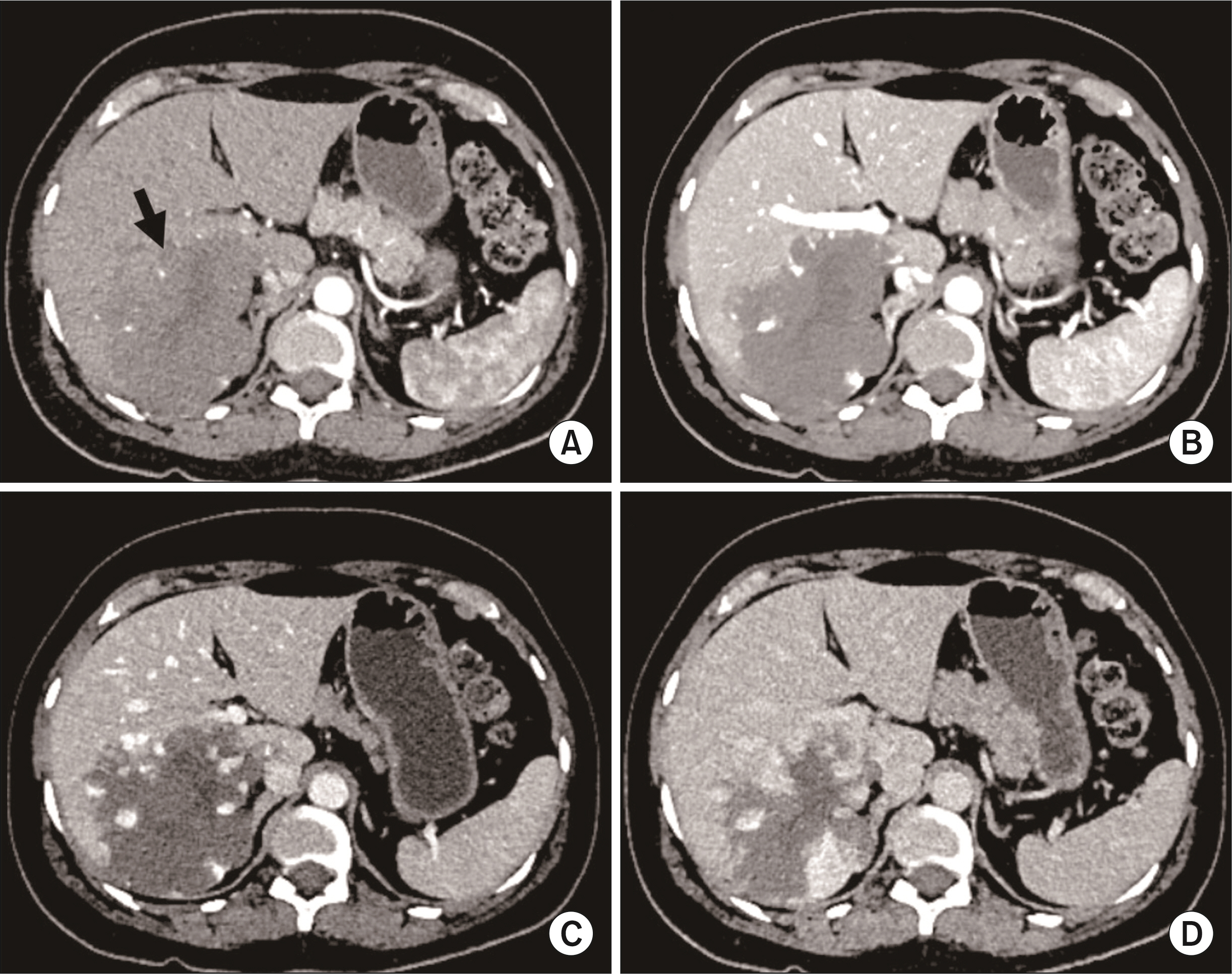
Fig. 1 Multiphasic contrast-enhanced computed tomography (CT) in a 52-year-old male patient showing a giant hepatic haemangioma (arrow in A). Axial images in (A) arterial, (B) portal venous, (C) hepatic venous, and (D) 3-minute delayed phases show typical peripheral discontinuous nodular enhancement with progressive centripetal filling of contrast. Note that the density of nodular contrast in the lesion matches with the aorta (blood pool).
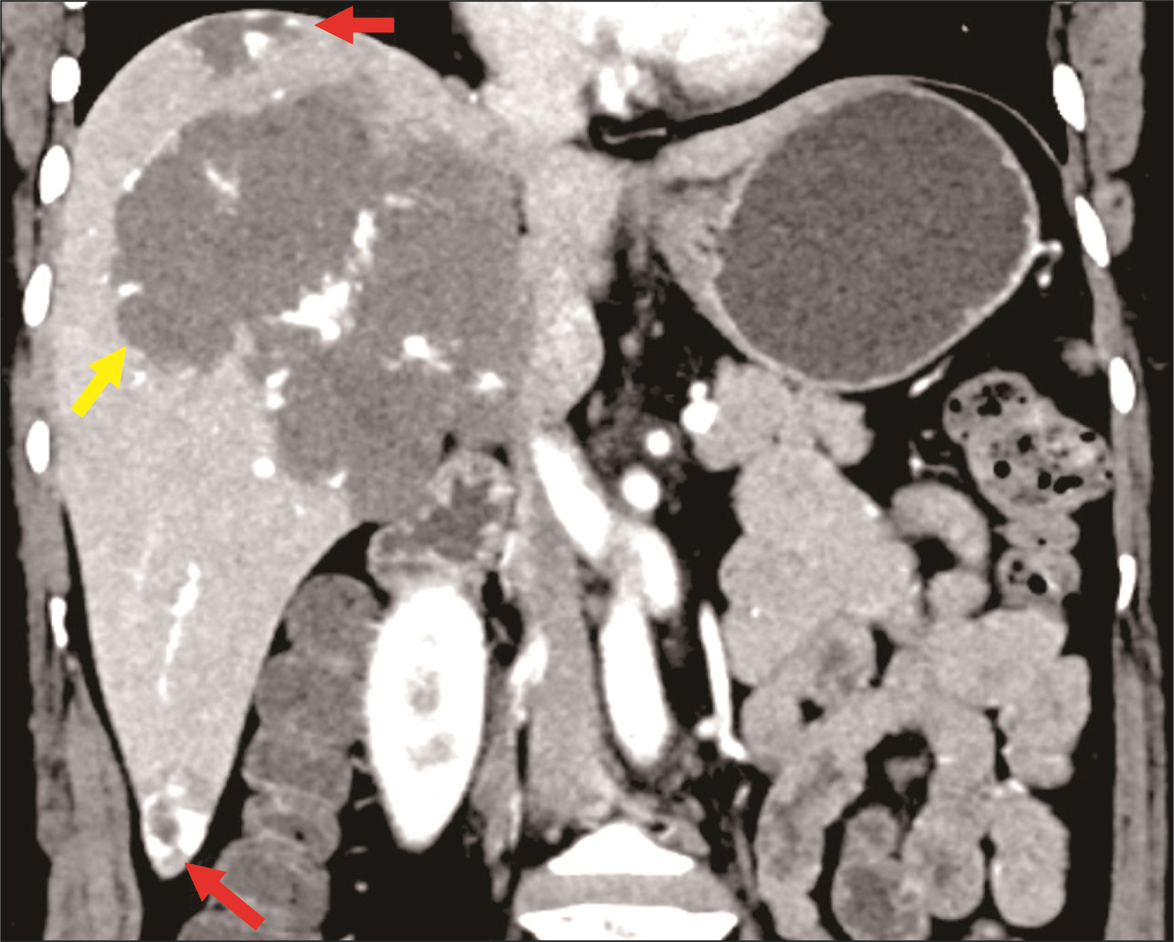
Fig. 2 Multiple hepatic haemangiomas on contrast-enhanced computed tomography (CT) in portal venous phase in a 42-year-old female. The coronal image shows two small haemangiomas (red arrows) and one giant haemangioma (yellow arrow).
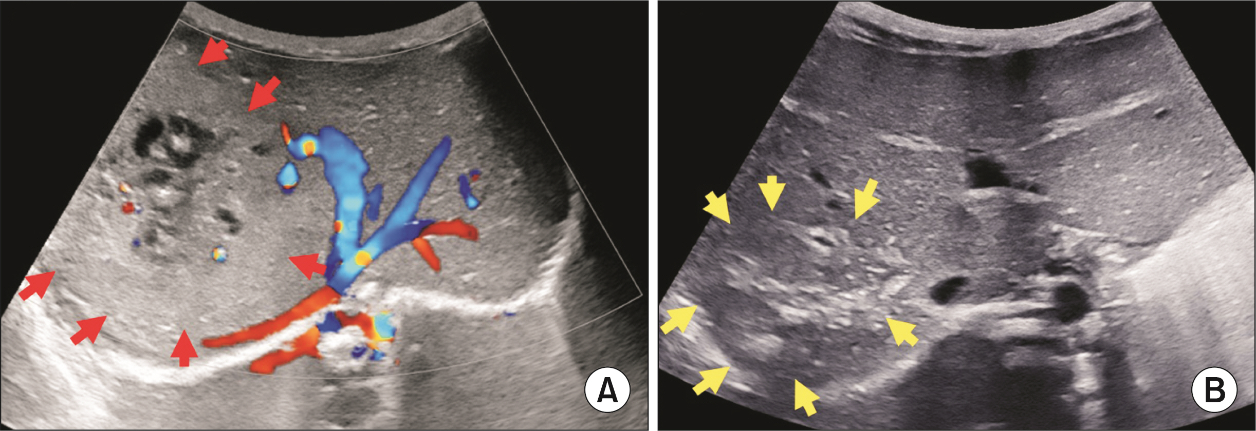
Fig. 3 Ultrasound images in an infant with congenital hepatic haemangioma. (A) Colour doppler image in a 19-day-old neonate shows a large hyperechoic mass with internal cystic areas and vascularity. Lesion is delineated by red arrows. Mass is causing splaying of hepatic veins. (B) Follow-up ultrasound at the age of 15 months shows a significant decrease in the size of the mass lesion with increased hyperechoic fibrous component. Mass is delineated by yellow arrows.
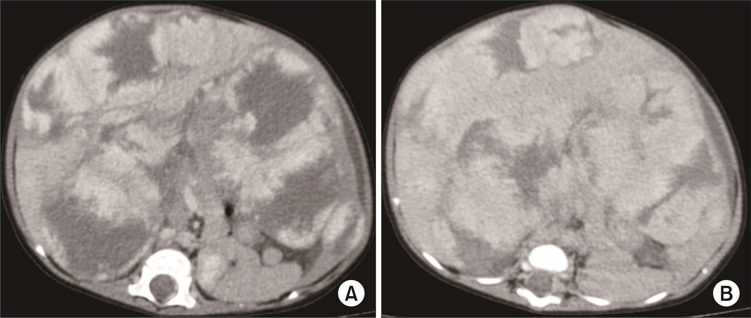
Fig. 4 Contrast-enhanced computed tomography (CT) images in a 28-day-old female neonate showing diffuse infantile hepatic haemangiomas who presented with abdominal distension. Axial images in (A) portal venous, (B) 3-minute delayed phases show typical peripheral nodular enhancement with progressive centripetal filling of contrast in delayed phase. Intervening normal liver parenchyma is seen between lesions.

Fig. 5 Multiphasic contrast-enhanced computed tomography (CT) in a 45-year-old male patient showing a large regenerative nodule in the background of Budd-Chiari syndrome. Axial images in (A) non-contrast, (B) arterial, (C) portal venous, and (D) hepatic venous phases show a well-defined nodule in segment II (red arrow). Nodule is iso-hypoattenuating in non-contrast CT (A), showing arterial enhancement (B) which homogeneously progresses in portal venous phase and becomes isoattenuating in hepatic venous phase (D) with the background parenchyma. Hepatomegaly, heterogeneous reticular enhancement of liver, splenomegaly, prominent azygous system, and ascites are features of Budd-Chiari syndrome seen in this case due to supra hepatic inferior vena cava (IVC) stenosis.

Fig. 6 Multiple hepatic epitheloid hemangioendotheliomas on multiphasic contrast-enhanced computed tomography (CT) (all axial images at three different levels) in a 35-year-old male patient. (A, B, C) Arterial phase, (D, E, F) venous phase, (G, H, I) 3-minute delayed phase images showing multiple peripheral subcapsular lesions with progressive enhancement. Lesions are causing subcapsular retraction (red arrows) leading to a nodular outline of the liver. Some lesions are coalescing to form confluent masses along the right lobe (yellow arrows in E).
Reference
-
1. Lerut J, Iesari S. 2018; Vascular tumours of the liver: a particular story. Transl Gastroenterol Hepatol. 3:62. DOI: 10.21037/tgh.2018.09.02. PMID: 30363746. PMCID: PMC6182012.
Article2. WHO Classification of Tumours Editorial Board. Soft tissue and bone tumours. Vol 3:5th ed. International Agency for Research on Cancer;2020.3. Bannoura S, Putra J. 2021; Primary malignant vascular tumors of the liver in children: angiosarcoma and epithelioid hemangioendothelioma. World J Gastrointest Oncol. 13:223–230. DOI: 10.4251/wjgo.v13.i4.223. PMID: 33889274. PMCID: PMC8040065.
Article4. Bridgewater J, Galle PR, Khan SA, Llovet JM, Park JW, Patel T, et al. 2014; Guidelines for the diagnosis and management of intrahepatic cholangiocarcinoma. J Hepatol. 60:1268–1289. DOI: 10.1016/j.jhep.2014.01.021. PMID: 24681130.
Article5. European Association for the Study of the Liver. 2018; EASL Clinical Practice Guidelines: management of hepatocellular carcinoma. J Hepatol. 69:182–236. DOI: 10.1016/j.jhep.2019.01.020. PMID: 29628281.6. Dasgupta R, Fishman SJ. 2014; ISSVA classification. Semin Pediatr Surg. 23:158161. DOI: 10.1053/j.sempedsurg.2014.06.016. PMID: 25241091.
Article7. Choi BY, Nguyen MH. 2005; The diagnosis and management of benign hepatic tumours. J Clin Gastroenterol. 39:401–412. DOI: 10.1097/01.mcg.0000159226.63037.a2. PMID: 15815209.8. Dokmak S, Ronot M. Jarnagin W, editor. 2023. Chapter 88A - Benign liver lesions. Blumgart’s Surgery of the Liver, Biliary tract and Pancreas. Elsevier Inc.;7th ed. p. 1181–1202.9. Ehman EC, Torbenson MS, Wells ML, Welch BT, Thompson SM, Garg I, et al. 2018; Hepatic tumours of vascular origin: imaging appearances. Abdom Radiol (NY). 43:1978–1990. DOI: 10.1007/s00261-017-1401-3. PMID: 29159525.10. Ros PR, Erturk SM. Gore RM, Levine MS, editors. 2021. Benign Tumours of the Liver. Textbook of Gastrointestinal Radiology. 5th ed. Elsevier Inc.;p. 767–787. DOI: 10.1159/000060935.11. Thampy R, Elsayes KM, Menias CO, Pickhardt PJ, Kang HC, Deshmukh SP, et al. 2017; Imaging features of rare mesenychmal liver tumours: beyond haemangiomas. Br J Radiol. 90:20170373. DOI: 10.1259/bjr.20170373. PMID: 28766950. PMCID: PMC5963373.
Article12. Miyaki D, Aikata H, Waki K, Murakami E, Hashimoto Y, Nagaoki Y, et al. 2011; [Significant regression of a cavernous hepatic haemangioma to a sclerosed haemangioma over 12 years: a case study]. Nihon Shokakibyo Gakkai Zasshi. 108:954–961. Japanese.13. Yoo BR, Han HY, Choi SY, Kim JH. 2014; Giant cavernous haemangioma coexistent with diffuse hepatic haemangiomatosis presenting as portal vein thrombosis and hepatic lobar atrophy. Ultrasonography. 33:65–70. DOI: 10.14366/usg.13003. PMID: 24936497. PMCID: PMC4058974.
Article14. Conter RL, Longmire WP Jr. 1988; Recurrent hepatic haemangiomas. Possible association with estrogen therapy. Ann Surg. 207:115–119. DOI: 10.1097/00000658-198802000-00001. PMID: 2829759. PMCID: PMC1493383.15. Feurle GE. 1990; Arteriovenous shunting and cholestasis in hepatic haemangiomatosis associated with metoclopramide. Gastroenterology. 99:258–262. DOI: 10.1016/0016-5085(90)91256-6. PMID: 2344931.
Article16. Maeda E, Akahane M, Watadani T, Yoshioka N, Goto A, Sugawara Y, et al. 2007; Isolated hepatic haemangiomatosis in adults: report of two cases and review of the literature. Eur J Radiol Extra. 61:9–14. DOI: 10.1016/j.ejrex.2006.10.007.
Article17. Chung EM, Lattin GE Jr, Cube R, Lewis RB, Marichal-Hernández C, Shawhan R, et al. 2011; From the archives of the AFIP: pediatric liver masses: radiologic-pathologic correlation. Part 2. Malignant tumours. Radiographics. 31:483–507. DOI: 10.1148/rg.312105201. PMID: 21415193.18. Hsi Dickie B, Fishman SJ, Azizkhan RG. 2014; Hepatic vascular tumours. Semin Pediatr Surg. 23:168–172. DOI: 10.1053/j.sempedsurg.2014.06.018. PMID: 25241093.19. Bisceglie AMD. Feldman M, Friedman LS, Brandt LJ, editors. 2021. Chapter 96 - Hepatic Tumours and Cysts. Sleisenger and Fordtran’s Gastrointestinal and Liver Disease. 11th ed. Elsevier Inc.;p. 1509–1532.20. Paradis V. Jarnagin W, editor. 2023. Chapter 87 - Tumours of the liver: Pathologic aspects. Blumgart’s Surgery of the Liver, Biliary tract and Pancreas. 7th ed. Elsevier Inc.;p. 1152–1180.21. Kristidis P, de Silva M, Howman-Giles R, Gaskin KJ. 1991; Infantile hepatic haemangioma: investigation and treatment. J Paediatr Child Health. 27:57–61. DOI: 10.1111/j.1440-1754.1991.tb00348.x. PMID: 2043394.22. Mo JQ, Dimashkieh HH, Bove KE. 2004; GLUT1 endothelial reactivity distinguishes hepatic infantile haemangioma from congenital hepatic vascular malformation with associated capillary proliferation. Hum Pathol. 35:200–209. DOI: 10.1016/j.humpath.2003.09.017. PMID: 14991538.23. Shastri S, Dubinsky MC, Fred Poordad F, Vasiliauskas EA, Geller SA. 2004; Early nodular hyperplasia of the liver occurring with inflammatory bowel diseases in association with thioguanine therapy. Arch Pathol Lab Med. 128:49–53. DOI: 10.5858/2004-128-49-ENHOTL. PMID: 14692812.
Article24. WHO Classification of Tumours Editorial Board. Digestive system tumours. 5th ed. International Agency for Research on Cancer;2019. DOI: 10.5858/2004-128-49-enhotl.25. Restrepo CS, Martínez S, Lemos JA, Carrillo JA, Lemos DF, Ojeda P, et al. 2006; Imaging manifestations of Kaposi sarcoma. Radiographics. 26:1169–1185. DOI: 10.1148/rg.264055129. PMID: 16844940.
Article26. Addula D, Das CJ, Kundra V. 2021; Imaging of Kaposi sarcoma. Abdom Radiol (NY). 46:5297–5306. DOI: 10.1007/s00261-021-03205-6. PMID: 34255129. PMCID: PMC8502139.
Article27. Nie P, Wu J, Wang H, Zhou R, Sun L, Chen J, et al. 2019; Primary hepatic perivascular epithelioid cell tumours: imaging findings with histopathological correlation. Cancer Imaging. 19:32. DOI: 10.1186/s40644-019-0212-x. PMID: 31171030. PMCID: PMC6555711.28. Shen HQ, Chen DF, Sun XH, Li X, Xu J, Hu XB, et al. 2013; MRI diagnosis of perivascular epithelioid cell tumour (PEComa) of the liver. Rom J Morphol Embryol. 54:643–647.29. Maebayashi T, Abe K, Aizawa T, Sakaguchi M, Ishibashi N, Abe O, et al. 2015; Improving recognition of hepatic perivascular epithelioid cell tumour: case report and literature review. World J Gastroenterol. 21:5432–5441. DOI: 10.3748/wjg.v21.i17.5432. PMID: 25954119. PMCID: PMC4419086.
Article30. Folpe AL, Kwiatkowski DJ. 2010; Perivascular epithelioid cell neoplasms: pathology and pathogenesis. Hum Pathol. 41:1–15. DOI: 10.1016/j.humpath.2009.05.011. PMID: 19604538.
Article31. Lazăr DC, Avram MF, Romoșan I, Văcariu V, Goldiș A, Cornianu M. 2019; Malignanthepatic vascular tumours in adults: characteristics, diagnostic difficulties and currentmanagement. World J Clin Oncol. 10:110–135. DOI: 10.5306/wjco.v10.i3.110. PMID: 30949442. PMCID: PMC6441663.
Article32. Yugawa K, Yoshizumi T, Mano Y, Kurihara T, Yoshiya S, Takeishi K, et al. 2019; Solitary fibrous tumour in the liver: case report and literature review. Surg Case Rep. 5:68. DOI: 10.1186/s40792-019-0625-6. PMID: 31020464. PMCID: PMC6482201.33. Chan G, Horton PJ, Thyssen S, Lamarche M, Nahal A, Hill DJ, et al. 2007; Malignant transformation of a solitary fibrous tumour of the liver and intractable hypoglycemia. J Hepatobiliary Pancreat Surg. 14:595–599. DOI: 10.1007/s00534-007-1210-0. PMID: 18040628.
Article34. Bejarano-González N, García-Borobia FJ, Romaguera-Monzonís A, García-Monforte N, Falcó-Fagés J, Bella-Cueto MR, et al. 2015; Solitary fibrous tumour of the liver. Case report and review of the literature. Rev Esp Enferm Dig. 107:633–639. DOI: 10.17235/reed.2015.3676/2014. PMID: 15221390.35. Adams J, Lodge JP, Parker D. 1999; Liver transplantation for metastatic hemangiopericytoma associated withhypoglycemia. Transplantation. 67:488–489. DOI: 10.1097/00007890-199902150-00027. PMID: 10030302.
Article36. Miller WJ, Dodd GD 3rd, Federle MP, Baron RL. 1992; Epithelioid hemangioendothelioma of the liver: imaging findings with pathologic correlation. AJR Am J Roentgenol. 159:53–57. DOI: 10.2214/ajr.159.1.1302463. PMID: 1302463.
Article37. Da Ines D, Petitcolin V, Joubert-Zakeyh J, Demeocq F, Garcier JM. 2010; Epithelioid hemangioendothelioma of the liver with metastatic coeliac lymph nodes in an 11-year-old boy. Pediatr Radiol. 40:1293–1296. DOI: 10.1007/s00247-009-1532-y. PMID: 20112013.
Article38. Ros PR, Erturk SM. Gore RM, Levine MS, editors. 2021. Malignant Tumours of the Liver. Textbook of Gastrointestinal Radiology. 5th ed. Elsevier Inc.;p. 788–818.39. Lyburn ID, Torreggiani WC, Harris AC, Zwirewich CV, Buckley AR, Davis JE, et al. 2003; Hepatic epithelioid hemangioendothelioma: sonographic, CT, and MR imaging appearances. AJR Am J Roentgenol. 180:1359–1364. DOI: 10.2214/ajr.180.5.1801359. PMID: 12704052.
Article40. Lau K, Massad M, Pollak C, Rubin C, Yeh J, Wang J, et al. 2011; Clinical patterns and outcome in epithelioid hemangioendothelioma with or without pulmonary involvement: insights from an internet registry in the study of a rare cancer. Chest. 140:1312–1318. DOI: 10.1378/chest.11-0039. PMID: 21546438.
Article41. Flucke U, Vogels RJ, de Saint Aubain Somerhausen N, Creytens DH, Riedl RG, van Gorp JM, et al. 2014; Epithelioid hemangioendothelioma: clinicopathologic, immunhistochemical, and molecular genetic analysis of 39 cases. Diagn Pathol. 9:131. DOI: 10.1186/1746-1596-9-131. PMID: 24986479. PMCID: PMC4100035.
Article42. Doyle LA, Fletcher CD, Hornick JL. 2016; Nuclear expression of CAMTA1 distinguishes epithelioid hemangioendothelioma from histologic mimics. Am J Surg Pathol. 40:94–102. DOI: 10.1097/PAS.0000000000000511. PMID: 26414223.
Article43. Antonescu CR, Le Loarer F, Mosquera JM, Sboner A, Zhang L, Chen CL, et al. 2013; Novel YAP1-TFE3 fusion defines a distinct subset of epithelioid hemangioendothelioma. Genes Chromosomes Cancer. 52:775–784. DOI: 10.1002/gcc.22073. PMID: 23737213. PMCID: PMC4089994.
Article44. Levy DW, Rindsberg S, Friedman AC, Fishman EK, Ros PR, Radecki PD, et al. 1986; Thorotrast-induced hepatosplenic neoplasia: CT identification. AJR Am J Roentgenol. 146:997–1004. DOI: 10.2214/ajr.146.5.997. PMID: 3008544.
Article45. Zheng YW, Zhang XW, Zhang JL, Hui ZZ, Du WJ, Li RM, et al. 2014; Primary hepatic angiosarcoma and potential treatment options. J Gastroenterol Hepatol. 29:906–911. DOI: 10.1111/jgh.12506. PMID: 24372769.
Article46. Worawattanakul S, Semelka RC, Kelekis NL, Woosley JT. 1997; Angiosarcoma of the liver: MR imaging pre- and post-chemotherapy. Magn Reson Imaging. 15:613–617. DOI: 10.1016/S0730-725X(96)00393-1. PMID: 9254006.
Article47. Yasir S, Torbenson MS. 2019; Angiosarcoma of the liver: clinicopathologic features and morphologic patterns. Am J Surg Pathol. 43:581–590. DOI: 10.1097/PAS.0000000000001228. PMID: 30986799.48. Mehrabi A, Kashfi A, Fonouni H, Schemmer P, Schmied BM, Hallscheidt P, et al. 2006; Primary malignant hepatic epithelioid hemangioendothelioma: a comprehensive review of the literature with emphasis on the surgical therapy. Cancer. 107:2108–2121. DOI: 10.1002/cncr.22225. PMID: 17019735.
Article49. Li DB, Si XY, Wan T, Zhou YM. 2018; A pooled analysis of treatment and prognosis of hepatic angiosarcoma in adults. Hepatobiliary Pancreat Dis Int. 17:198–203. DOI: 10.1016/j.hbpd.2018.04.005. PMID: 29724676.
Article50. Omiyale AO, Carton J. 2018; Clinical and pathologic features of primary angiosarcoma of the kidney. Curr Urol Rep. 19:4. DOI: 10.1007/s11934-018-0755-6. PMID: 29383452.
Article51. Huang NC, Kuo YC, Chiang JC, Hung SY, Wang HM, Hung YM, et al. 2015; Hepatic angiosarcoma may have fair survival nowadays. Medicine (Baltimore). 94:e816. DOI: 10.1097/MD.0000000000000816. PMID: 25984668. PMCID: PMC4602568.
Article52. Goh IY, Mulholland P, Sokolova A, Liu C, Siriwardhane M. 2021; Hepatic small vessel neoplasm - a systematic review. Ann Med Surg (Lond). 72:103004. DOI: 10.1016/j.amsu.2021.103004. PMID: 34815856. PMCID: PMC8591473.
Article53. Gill RM, Buelow B, Mather C, Joseph NM, Alves V, Brunt EM, et al. 2016; Hepatic small vessel neoplasm, a rare infiltrative vascular neoplasm of uncertain malignant potential. Hum Pathol. 54:143–151. DOI: 10.1016/j.humpath.2016.03.018. PMID: 27090685. PMCID: PMC5242228.
Article54. Lewis S, Aljarallah B, Trivedi A, Thung SN. 2015; Magnetic resonance imaging of a small vessel hepatic haemangioma in a cirrhotic patient with histopathologic correlation. Clin Imaging. 39:702–706. DOI: 10.1016/j.clinimag.2015.02.007. PMID: 25748474.
Article55. Rangaswamy B, Minervini M, Tublin M, Sholosh B, Dasyam AK. 2019; Imaging and pathologic findings of hepatic small vessel haemangioma. Curr Probl Diagn Radiol. 48:626–628. DOI: 10.1067/j.cpradiol.2018.02.006. PMID: 29576414.56. Walcott-Sapp S, Tang E, Kakar S, Shen J, Hansen P. 2018; Resection of the largest reported hepatic small vessel neoplasm. Hum Pathol. 78:159–162. DOI: 10.1016/j.humpath.2018.01.013. PMID: 29366622.
Article57. Joseph NM, Brunt EM, Marginean C, Nalbantoglu I, Snover DC, Thung SN, et al. 2018; Frequent GNAQ and GNA14 mutations in hepatic small vessel neoplasm. Am J Surg Pathol. 42:1201–1207. DOI: 10.1097/PAS.0000000000001110. PMID: 29975248.
Article58. Yoon SS, Charny CK, Fong Y, Jarnagin WR, Schwartz LH, Blumgart LH, et al. 2003; Diagnosis, management, and outcomes of 115 patients with hepatic haemangioma. J Am Coll Surg. 197:392–402. DOI: 10.1016/S1072-7515(03)00420-4. PMID: 12946794.
Article59. Brouwers MA, Peeters PM, de Jong KP, Haagsma EB, Klompmaker IJ, Bijleveld CM, et al. 1997; Surgical treatment of giant haemangioma of the liver. Br J Surg. 84:314–316. DOI: 10.1046/j.1365-2168.1997.02534.x. PMID: 9117293.
Article60. Christison-Lagay ER, Burrows PE, Alomari A, Dubois J, Kozakewich HP, Lane TS, et al. 2007; Hepatic haemangiomas: subtype classification and development of a clinical practice algorithm and registry. J Pediatr Surg. 42:62–67. DOI: 10.1016/j.jpedsurg.2006.09.041. PMID: 17208542.61. Bajenaru N, Balaban V, Săvulescu F, Campeanu I, Patrascu T. Hepatic hemangioma -review-. J Med Life. 2015; 8 Spec Issue(Spec Issue):4–11. PMID: 26361504. PMCID: PMC4564031.62. Jia K, Gao Z, Li M, Yu C. 2022; Interventional treatments for hepatic hemangioma: A state-of-the-art review. J Interv Med. 5:6–9. DOI: 10.1016/j.jimed.2021.12.009. PMID: 35586280. PMCID: PMC8947984.63. Van Leer-Greenberg B, Kole A, Chawla S. 2017; Hepatic Kaposi sarcoma: A case report and review of the literature. World J Hepatol. 9:171–179. DOI: 10.4254/wjh.v9.i4.171. PMID: 28217255. PMCID: PMC5295157.
Article64. Arora M, Goldberg EM. 2010; Kaposi sarcoma involving the gastrointestinal tract. Gastroenterol Hepatol (N Y). 6:459–462. PMID: 20827371. PMCID: PMC2933764.65. Mulholland P, Goh IY, Sokolova A, Liu C, Siriwardhane M. 2021; Hepatic small vessel neoplasm case report: A surveillance conundrum. Int J Surg Case Rep. 81:105742. DOI: 10.1016/j.ijscr.2021.105742. PMID: 33743248. PMCID: PMC8010386.
Article66. Kumar A, Sharma B, Samant H. 2022. Liver Angiosarcoma [Internet]. StatPearls Publishing;Available from: https://www.ncbi.nlm.nih.gov/books/NBK538224/.67. Chen N, Yu AJS, Jung J. 2018; Primary hepatic angiosarcoma: a brief review of the literature. EMJ Hepatol. 6:64–71. DOI: 10.33590/emjhepatol/10314175.
Article68. Kou K, Chen YG, Zhou JP, Sun XD, Sun DW, Li SX, et al. 2020; Hepatic epithelioid hemangioendothelioma: Update on diagnosis and therapy. World J Clin Cases. 8:3978–3987. DOI: 10.12998/wjcc.v8.i18.3978. PMID: 33024754. PMCID: PMC7520791.69. Virarkar M, Saleh M, Diab R, Taggart M, Bhargava P, Bhosale P. 2020; Hepatic hemangioendothelioma: an update. World J Gastrointest Oncol. 12:248–266. DOI: 10.4251/wjgo.v12.i3.248. PMID: 32206176. PMCID: PMC7081107.
Article
- Full Text Links
-
- Actions
-
Cited
- CITED
-
- Close
- Share
-
- Similar articles
-
- The lymphatic vascular system in liver diseases: its role in ascites formation
- Hepatocellular Adenoma and Focal Nodular Hyperplasia
- Surgical management of hilar cholangiocarcinoma: Controversies and recommendations
- Contrast-enhanced ultrasonography of the liver using SonoVue
- Neuroendocrine tumors of the liver
