J Yeungnam Med Sci.
2023 Jan;40(1):109-111. 10.12701/jyms.2022.00626.
Right arm pain after strength training: ultrasound imaging for pectoralis major tendon strain
- Affiliations
-
- 1Department of Physical Medicine and Rehabilitation, Lo-Hsu Medical Foundation, Inc., Lotung Poh-Ai Hospital, Yilan, Taiwan
- 2Department of Physical Medicine and Rehabilitation, National Taiwan University Hospital and National Taiwan University College of Medicine, Taipei, Taiwan
- 3Department of Physical Medicine and Rehabilitation, Bei-Hu Branch of National Taiwan University Hospital, Taipei, Taiwan
- 4Center for Regional Anesthesia and Pain Medicine, Wang-Fang Hospital, Taipei Medical University, Taipei, Taiwan
- 5Department of Physical and Rehabilitation Medicine, Hacettepe University Medical School, Ankara, Turkey
- KMID: 2538794
- DOI: http://doi.org/10.12701/jyms.2022.00626
Figure
-
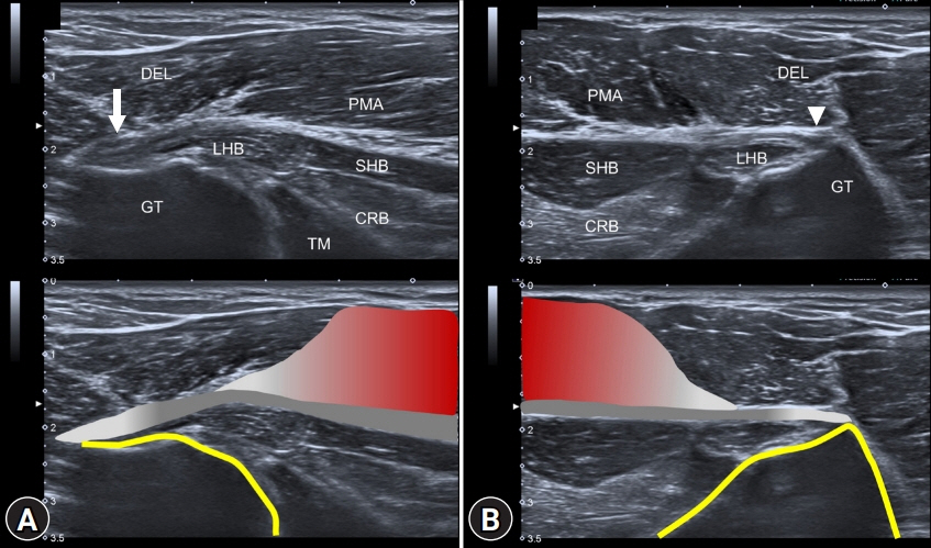
Fig. 1. (A) Ultrasound imaging of the right proximal region shows loss of fibrillar pattern and increased thickness of the pectoralis major tendon (arrow) near its insertion on the humerus. (B) The pectoralis major tendon (arrowhead) on the unaffected side has normal thickness and fibrillar pattern. CRB, coracobrachialis muscle; DEL, deltoid muscle; GT, greater tubercle; LHB, long head of the biceps muscle; SHB, short head of the biceps muscle; TM, teres major muscle; PMA, pectoralis major muscle.
Reference
-
References
1. Chang KV, Lin CP, Lin CS, Wu WT, Özçakar L. A novel approach for ultrasound guided axillary nerve block: the inferior axilla technique. Med Ultrason. 2017; 19:457–61.2. Butt U, Mehta S, Funk L, Monga P. Pectoralis major ruptures: a review of current management. J Shoulder Elbow Surg. 2015; 24:655–62.3. Fung L, Wong B, Ravichandiran K, Agur A, Rindlisbacher T, Elmaraghy A. Three-dimensional study of pectoralis major muscle and tendon architecture. Clin Anat. 2009; 22:500–8.4. ElMaraghy AW, Devereaux MW. A systematic review and comprehensive classification of pectoralis major tears. J Shoulder Elbow Surg. 2012; 21:412–22.5. Chiavaras MM, Jacobson JA, Smith J, Dahm DL. Pectoralis major tears: anatomy, classification, and diagnosis with ultrasound and MR imaging. Skeletal Radiol. 2015; 44:157–64.6. Petilon J, Carr DR, Sekiya JK, Unger DV. Pectoralis major muscle injuries: evaluation and management. J Am Acad Orthop Surg. 2005; 13:59–68.7. Chang KV, Wur WT, Özçakar L. Ultrasound imaging and rehabilitation of muscle disorders: part 1. Traumatic Injuries. Am J Phys Med Rehabil. 2019; 98:1133–41.8. Lee SJ, Jacobson JA, Kim SM, Fessell D, Jiang Y, Girish G, et al. Distal pectoralis major tears: sonographic characterization and potential diagnostic pitfalls. J Ultrasound Med. 2013; 32:2075–81.
- Full Text Links
-
- Actions
-
Cited
- CITED
-
- Close
- Share
-
- Similar articles
-
- Rupture of the Pectoralis Major Muscle during Exercise
- Accessory Tendon of Biceps Brachii Originated from Pectoralis Major
- Pectoralis Major Tendon Transfer for Refractory Winged Scapula: A Case Report
- Avulsion of Pectoralis Major Tendon: A Case Report
- RECONSTRUCTION OF A "THROUGH-AND-THROUGH" DEFECT OF BUCCAL CHEEK WITH BILOBULAR PECTORALIS MAJOR MYOCUTANEOUS ISLAND FLAP: REPORT OF A CASE & COMPARISON WITH A CONVENTIONAL PECTORALIS MAJOR MYOCUTANEOUS FLAP
